44 label the photomicrograph
A & P lab test 4 Flashcards | Quizlet Select all that apply. Label these structures of the upper respiratory system. Correctly label the components of the upper respiratory tract. Label the anterior view of the lower respiratory tract based on the hints if provided. Correctly label the components of the lungs. Correctly label the components of the pulmonary alveoli. Label The Photomicrograph Based On The Hints Provided. Capsule - 33 ... Label the photomicrograph based on the hints provided. Place each of the following lymphatic structures in the correct category based on their location. Searchable by topic and provided in ms word format, as well as in launchpad and diploma, the assessment bank offers a high level of flexibility.
1 2 label the photomicrograph of a transverse section 1 2 Label the photomicrograph of a transverse section of the spinal cord in Figure 17.5 Figure 17.5. LAB ACTIVITY 3: Transverse Section of Spinal Cord Identify the spinal cord structures in Figures 17.3 Figures 17.3 and 17.4 17.4 on a transverse section model or chart of the spinal cord, or use the search text box in Real Anatomy Real Anatomy (Nervous) to find these structures.

Label the photomicrograph
Label The Photomicrograph Of The Lung : 4 Chloro Dl Phenylalanine ... A photomicrograph of lung sections from (a) a control group showing normal epithelization of bronchi and bronchioles with normal alveoli and . Label The Photomicrograph Of The Lung : 4 Chloro Dl Phenylalanine Protects Against Monocrotaline Induced Pulmonary Vascular Remodeling And Lung Inflammation. The lower respiratory system., put the ... Endocrine System APR Module 8 Flashcards - Quizlet Label the photomicrograph based on the hints provided. (suprarenal gland) Label each of the following histology slides by dragging the histology slide of the gland under the correct name. Label The Photomicrograph Of Compact Bone. / Tik Ta Lk Aitee Umoffong ... Free answer to label the photomicrograph of compact bone. These bones do everything from protecting vital organs to giving muscles and nerves an anchor. (b) in this micrograph of the osteon, you can clearly see the concentric . The walls of the diaphysis are composed of dense and hard compact bone.
Label the photomicrograph. Label the photomicrograph based on the hints provided. the condition is identified by determining the level of oxygen in the blood sample obtained from an artery. it can also be predicted by determining the oxygen saturation of the blood with the application of a pulse oximeter. the normal arterial oxygen is about 75 to 100 mm hg. the values underneath 60 mm hg is generally considered with the ... Label the photomicrograph in figure 128 figure 128 - Course Hero Label the photomicrograph in Figure 12.8Figure 12.8. LAB ACTIVITY 3: Neuromuscular Junction Examine a prepared microscope slide of the neuromuscular junction and identify the structures listed in Figure 12.8Figure 12.8. Label the photomicrograph based on the hints provided. Pancreas ... Label the photomicrograph based on the hints provided. Pancreas Pancreatic islet Exocrine portion Pancreas Exocrine portion Intralobular duct Venule Pancreatic islet Arteriole Mar 29 2022 11:34 AM Solved > Question 31 points Label the photomicrograph of ... - ScholarOn Question : Question 31 points Label the photomicrograph of thin skin. Hair Follicle : 391984. Question. 31 points Label the photomicrograph of thin skin. Hair Follicle Hair Dermis Sebaceous gland Duct of sebaceous gland Reset zoom. Solution. 5 (1 Ratings ) Solved. Biology 2 Years Ago 68 Views.
[Solved] Please see an attachment for details | Course Hero Label the photomicrograph based on the hints provided. Medulla Capillary Zona fasciculara Suprarenal gland Zona reticularis... Biology Science Anatomy BISC 106. ... Make sure you label every questions you answers so i could be easy to follow. organize is key Part A 1. Define anatomy a. Q: ... Label The Photomicrograph Based On The Hints Provided / Endocrine Lab ... Label The Photomicrograph Based On The Hints Provided / Endocrine Lab Flashcards Quizlet. Spleen capsule capsule white pulp. Can be reproduced based on the information provided in the manuscript. A study based on observation and interview with individuals that uses inductive. Medulla capillary zona fasciculara suprarenal gland zona reticularis. Label The Photomicrograph Of The Sebaceous Gland : Histochemical ... 1 answer to label the photomicrograph of thin skin. If the gland become blocked, the sebum can be forced out into the dermis, where it elicits an inflammatory response. Photomicrograph of prepuce in golden jackal, (d) dermis, (sc) sebaceous gland, (sw) sweat gland, (g) guard hair follicle. fuiadinda64 April 01, 2022 URL Print Email Anatomy and Physiology Homework Chapter 6 Flashcards - Quizlet Label the photomicrograph of thick skin.-Stratum corneum-Stratum granulosum-Stratum spinosum-Stratum basale-Epidermis-Dermis-Stratum lucidum-Epidermis-Stratum corneum-Stratum lucidum-Stratum granulosum-Stratum spinosum-Stratum basale-Dermis Explanation: Thick skin is located on the palms and soles. Refer to APR 3.0 for further information.
Label The Photomicrograph Of Thin Skin Quizlet - Skin Labeling Review ... Label the photomicrograph of thin skin. Label the photomicrograph of thick skin. D) stratum corneum has fewer layers in. Start studying photomicrographs of skin (thin skin). Learn vocabulary, terms, and more with flashcards, games, and other study tools. C) contains more sweat glands than thin skin. Label the photomicrograph of thin skin. Label The Photomicrograph Using The Hints Provided / The Fate And ... Label The Photomicrograph Using The Hints Provided / The Fate And Toxicity Of Raman Active Silica Gold Nanoparticles In Mice. Correctly label the following anatomical parts of a kidney. Correctly label the following … Contribute to cth/sdcg development by creating an account on github. Label The Photomicrograph Using The Hints Provided : Pollen Microscope ... Photographic images can be taken through the microscope (photomicrography) by attaching a camera on to the vertical tube of the microscope's trinocular head label the photomicrograph. Using highly adherent human cervical tumor (hela) cells as a model. Routine stains are those used. Agranulocytes (includes lymphocytes and monocytes). Endocrine Practice for Lab Exam 1 .docx - Endocrine Practice for Lab ... Endocrine Practice for Lab Exam 1 1. Label the photomicrograph based on the hints provided. a. 2. Label each of the following histology slides by dragging the histology slide of the gland under the correct name. a.
![HLS [ Cartilage and Bone and Bone Histogenesis, compact bone] LOW MAG ...](http://www.bu.edu/histology/i/02701loa.jpg)
HLS [ Cartilage and Bone and Bone Histogenesis, compact bone] LOW MAG ...
Practical 2 Flashcards | Quizlet label each line on the pressure graph below as representing the aorta, left atrium, or left ventricle. identify the specific region on the graph associated with each phase of the cardiac cycle listed. click on the region of the ECG image below that aligns with the electrical changes related to atrial repolarization

Plant Cell Elodea Isotonic Solution Shows Cells Chloroplasts 250x At ...
Label The Photomicrograph Based On The Hints Provided. Zona Fasciculata ... Label The Photomicrograph Based On The Hints Provided. Zona Fasciculata / Repub Eur Nl. Capsule zona glomerulosa zona fasciculata capillaries suprarenal gland fascicle of cells . Label the photomicrograph based on the hints provided. Cortical sinus cortex medullary sinus secondary nodule primary nodule capsule lymph node medulla .
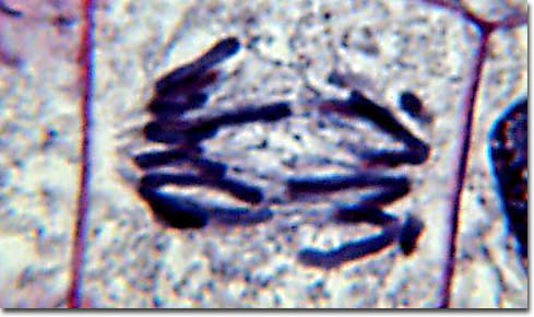
Molecular Expressions Photo Gallery: Mitosis - Early Anaphase
Ch. 22 Assessment Flashcards | Quizlet Label the structures in the photomicrograph based on the hints provided. List the correct order of lymphatic flow through a lymph node. 1. Afferent lymphatic vessel 2. Subcapsular sinus of the cortex 3. Sinuses of cortex and medulla 4. Efferent lymphatic vessel Put the following events into the correct order. 1.
PPT - Lab Exercise Classification of Tissues: Epithelial Tissue ...
Label The Photomicrograph Of Compact Bone. / Tik Ta Lk Aitee Umoffong ... Free answer to label the photomicrograph of compact bone. These bones do everything from protecting vital organs to giving muscles and nerves an anchor. (b) in this micrograph of the osteon, you can clearly see the concentric . The walls of the diaphysis are composed of dense and hard compact bone.
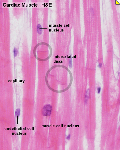
HM Practical - Cardiac Histology - Embryology
Endocrine System APR Module 8 Flashcards - Quizlet Label the photomicrograph based on the hints provided. (suprarenal gland) Label each of the following histology slides by dragging the histology slide of the gland under the correct name.

Pancreas - Human Physiology - 78 Steps Health
Label The Photomicrograph Of The Lung : 4 Chloro Dl Phenylalanine ... A photomicrograph of lung sections from (a) a control group showing normal epithelization of bronchi and bronchioles with normal alveoli and . Label The Photomicrograph Of The Lung : 4 Chloro Dl Phenylalanine Protects Against Monocrotaline Induced Pulmonary Vascular Remodeling And Lung Inflammation. The lower respiratory system., put the ...

Pseudostratified Ciliated Columnar Epithelium Shows Cilia Ciliated ...

Solved: The Histogram Shows The Amounts (in $) Of A Sample... | Chegg.com

Anatomy and Physiology 2 Eportfolio: Objective 79: Mommy what's that

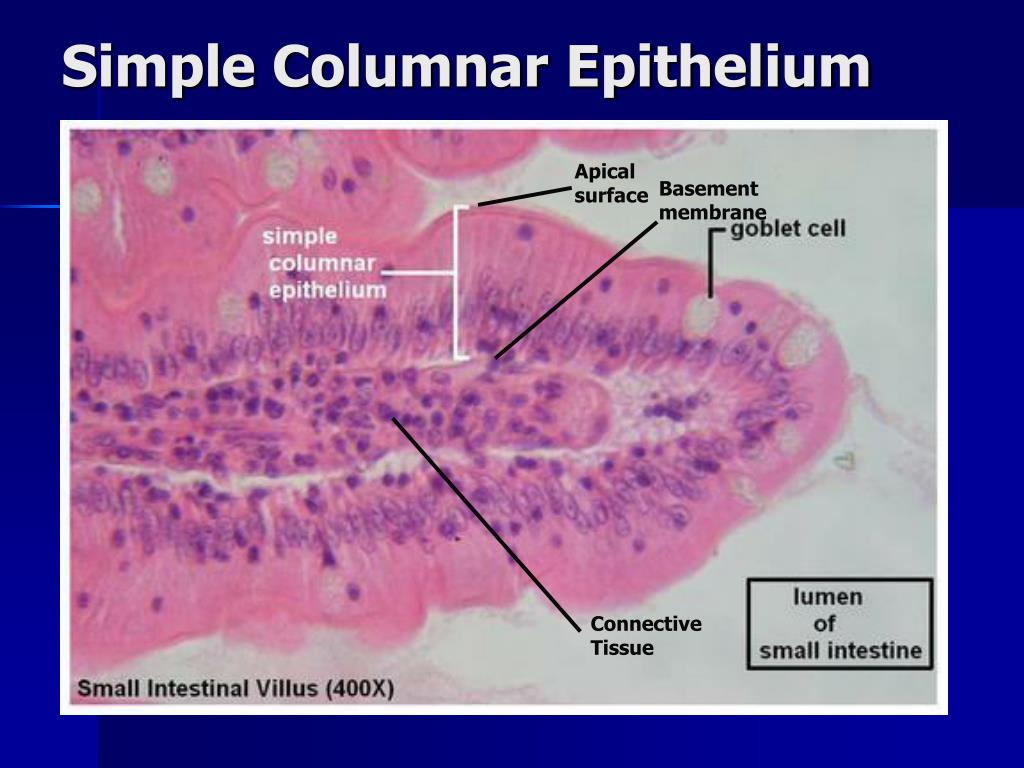
Photomicrograph Of Villus Of The Ileum Showing Simple Columnar ...
Post a Comment for "44 label the photomicrograph"