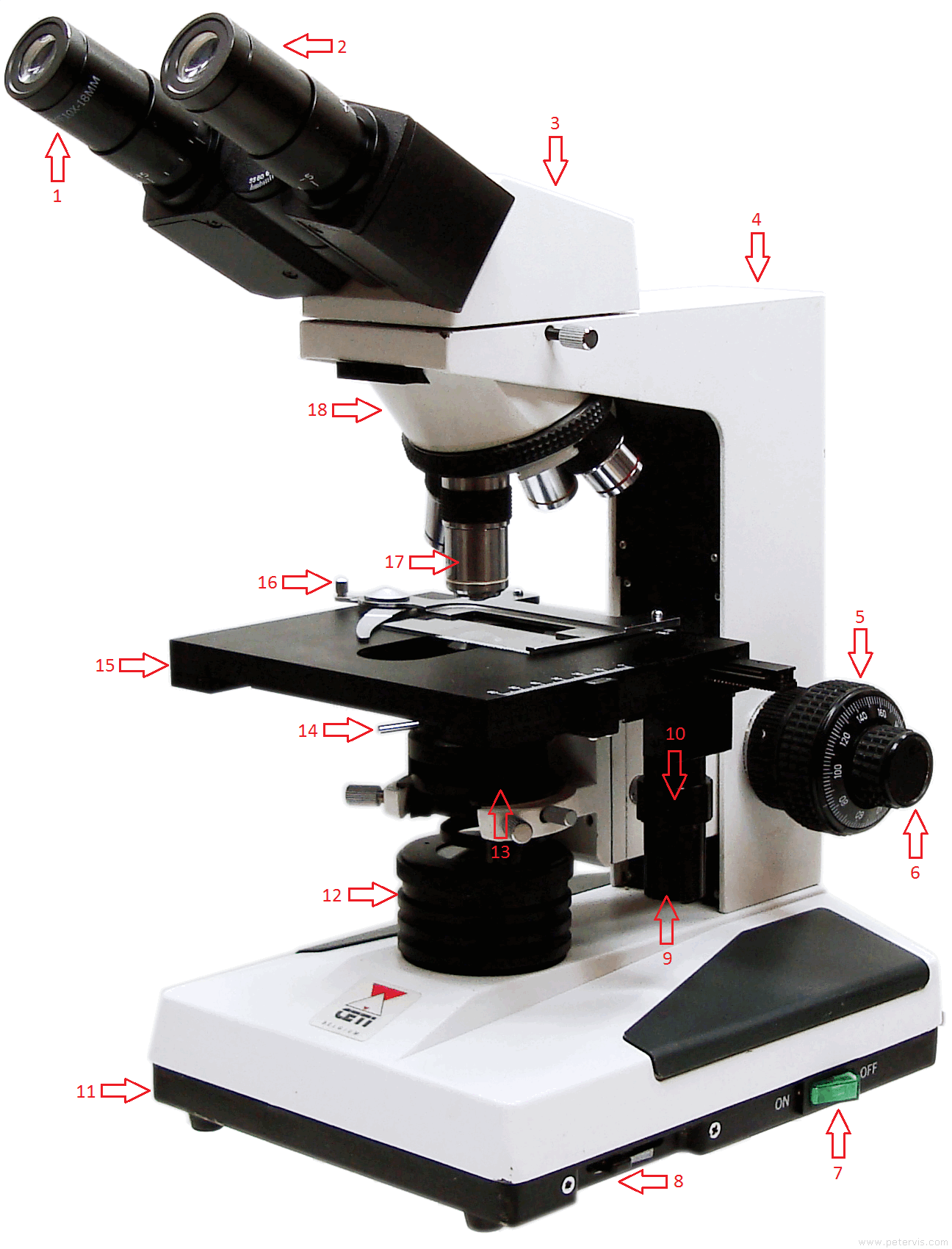
45 labelled diagram of a binocular microscope
en.wikipedia.org › wiki › EyeEye - Wikipedia Complex eyes distinguish shapes and colours.The visual fields of many organisms, especially predators, involve large areas of binocular vision for depth perception.In other organisms, particularly prey animals, eyes are located to maximise the field of view, such as in rabbits and horses, which have monocular vision. en.wikipedia.org › wiki › Parallax_barrierParallax barrier - Wikipedia The principle of the parallax barrier was independently invented by Auguste Berthier, who published an article on stereoscopic pictures including his new idea illustrated with a diagram and pictures with purposely exaggerated dimensions of the interlaced image strips, and by Frederic E. Ives, who made and exhibited a functional autostereoscopic image in 1901.
Microscope Parts, Function, & Labeled Diagram - slidingmotion Microscope parts labeled diagram gives us all the information about its parts and their position in the microscope. Microscope Parts Labeled Diagram The principle of the Microscope gives you an exact reason to use it. It works on the 3 principles. Magnification Resolving Power Numerical Aperture. Parts of Microscope Head Base Arm Eyepiece Lens

Labelled diagram of a binocular microscope
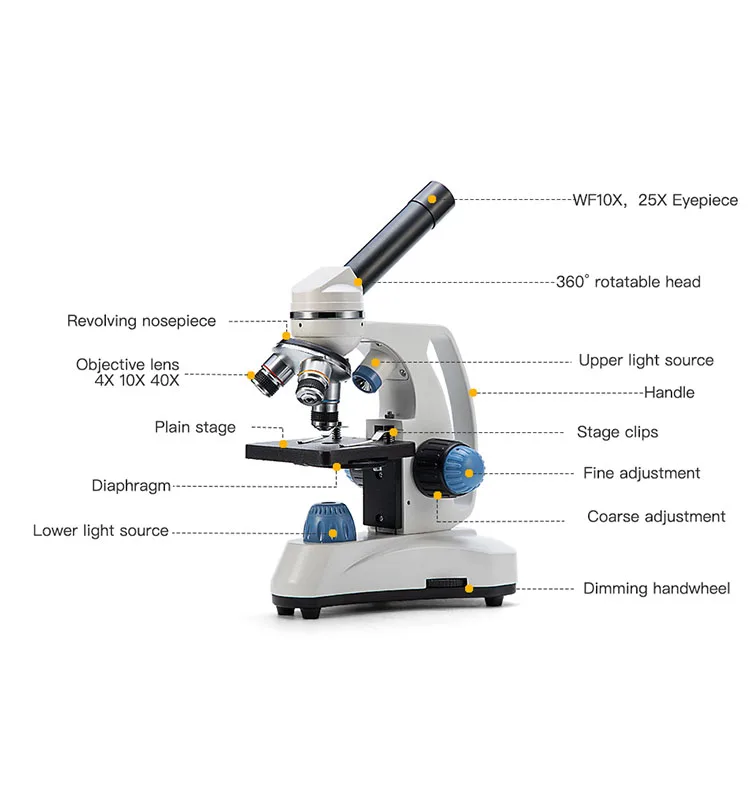
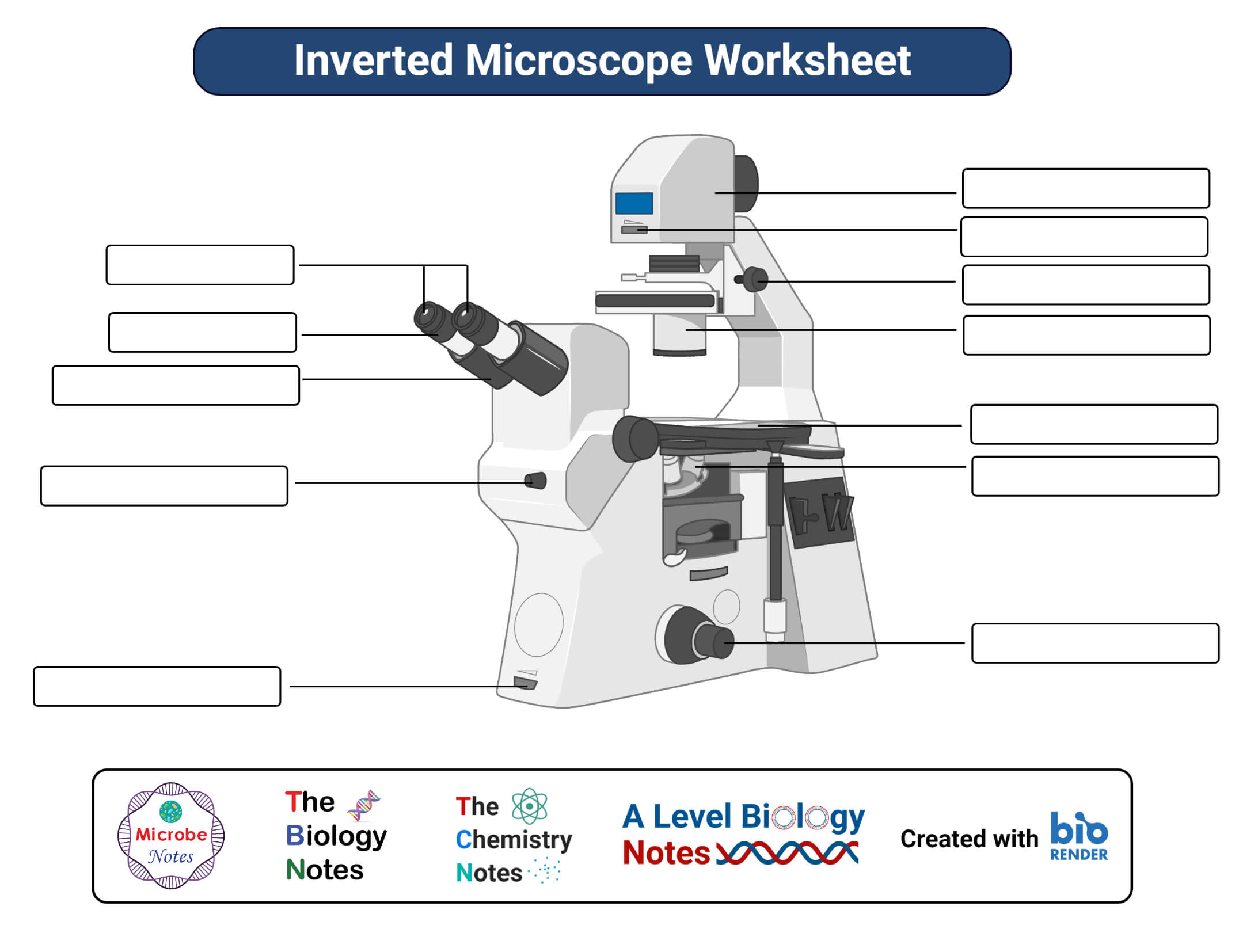
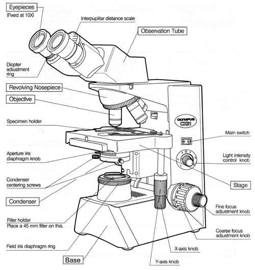
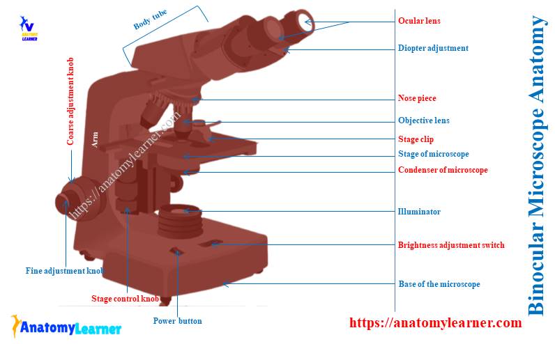
Parts of a microscope with functions and labeled diagram - Microbe Notes Q. List down the 18 parts of a Microscope. 1. Ocular Lens (Eye Piece) 2. Diopter Adjustment 3. Head 4. Nose Piece 5. Objective Lens 6. Arm (Carrying Handle) 7. Mechanical Stage 8. Stage Clip 9. Aperture 10. Diaphragm 11. Condenser 12. Coarse Adjustment 13. Fine Adjustment 14. Illuminator (Light Source) 15. Stage Controls 16. Base 17. Microscope Labeling Diagram | Quizlet Unit 2 Lesson 5 - Punnett Squares and Pedigrees. 4 terms. PGFry210. Unit 2 Lesson 4 - Heredity. 9 terms. PGFry210. Upgrade to remove ads. Only $2.99/month. Binocular Microscope Anatomy - Parts and Functions with a Labeled Diagram Now, I will discuss the details anatomy of the light compound microscope with the labeled diagram. Why it is called binocular: because it has two ocular lenses or an eyepiece on the head that attaches to the objective lens, this ocular lens magnifies the image produced by the objective lens. Binocular microscope parts and functions
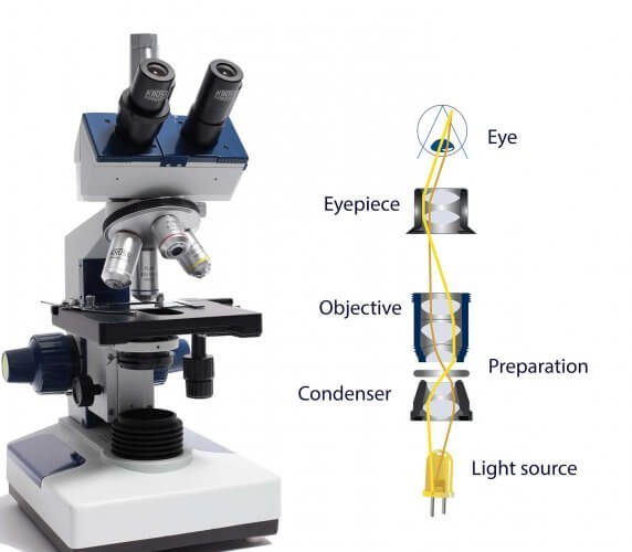
Labelled diagram of a binocular microscope. › exams › microscopeMicroscope: Types of Microscope, Parts, Uses, Diagram - Embibe Jul 19, 2022 · The stereo microscope, also called a dissecting microscope, provides magnification of up to \(300\) times. These binocular microscopes are used to look at opaque objects or objects that are too large to be viewed with a compound microscope since they do not require a slide preparation. Microscope Parts and Functions First, the purpose of a microscope is to magnify a small object or to magnify the fine details of a larger object in order to examine minute specimens that cannot be seen by the naked eye. Here are the important compound microscope parts... Eyepiece: The lens the viewer looks through to see the specimen. China Manufacture of Monocular Microscope Labelled Diagram, Monocular ... China Monocular Microscope Labelled Diagram manufacture, a number of high-quality Monocular Microscope Labelled Diagram sources of information for you to choose. Inquiry Basket ( 0) English; ... Multipurpose binocular infinite biology microscope. Monocular Microscope With Low Position Coaxial Coarse. Students Using Monocular Biological Microscope. What Are the Parts and Functions of a Binocular Microscope? Micah Elizabeth Scott/CC-BY-2.. The parts of a binocular microscope are the eye piece (ocular), mechanical stage, nose piece, objective lenses, condenser, lamp, microscope tube and prisms. Each part plays an important role in the microscope's function. Eye piece (ocular): The dual binocular eye piece contains the microscope's lenses and ...
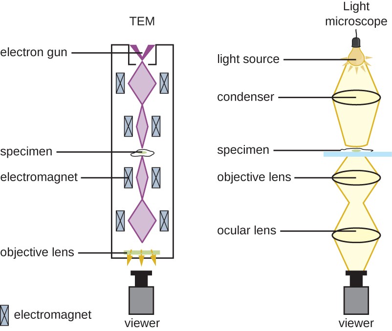
PaRTS of a BINOCULAR MICROSCOPE Flashcards | Quizlet arm one hand should grasp this part when carrying the microscope coarse adjustment knob this part is used to move the body up and down closer to the specimen fine adjustment used for fine, detailed focusing of the microscope light control controls the intensity of the light base one hand should be under this part when microscope is carried Monocular Microscope Labelled Diagram Exporters, Order Monocular ... Monocular Microscope Labelled Diagram - manufacturer, factory, supplier from China. Multi-Purpose Monocular Biological Microscope. Monocular / Binocular Biological Microscope With Low Position Coaxial Coarse en.wikipedia.org › wiki › Optical_microscopeOptical microscope - Wikipedia The optical microscope, also referred to as a light microscope, is a type of microscope that commonly uses visible light and a system of lenses to generate magnified images of small objects. Optical microscopes are the oldest design of microscope and were possibly invented in their present compound form in the 17th century. Diagram and labels of a monocular microscope? - Answers A monocular microscope has only one eyepiece while a binocular microscope has two eyepieces with different lenses. Binocular microscopes are more popular today than the monocular microscope for ...
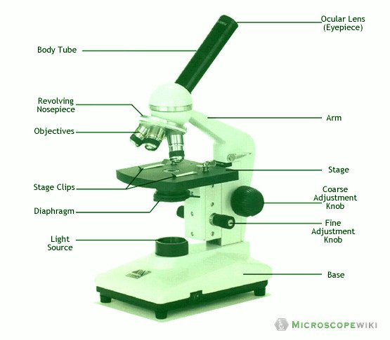
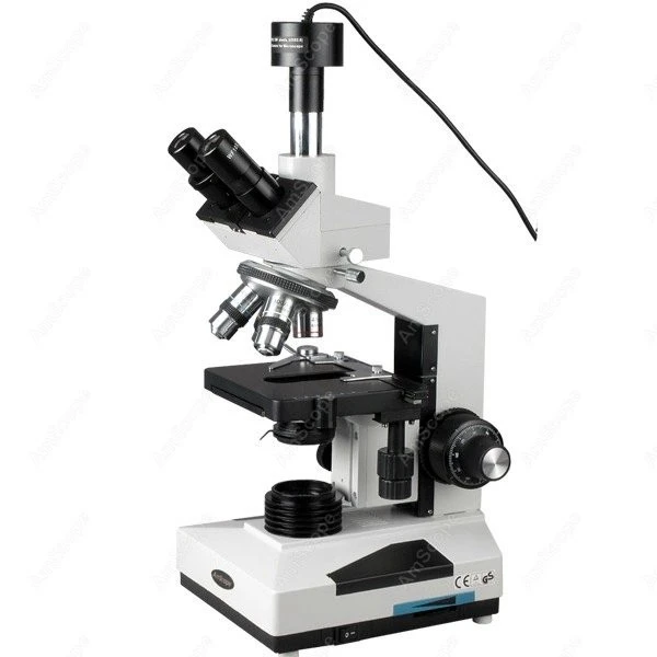
40+ Labelled Binocular Microscope Gif - Hortensia is Great The part that is looked through at the top of the compound microscope. Labeling the parts of the microscope. Sep 07, 2018 · labeled well labelled labeled binocular compound microscope. This activity has been designed for use in homes and schools. An ocular lenses and objective lenses. Draw a labelled ray diagram of a compound microscope and ... Diagrams of binocular microscope with labels? - Answers Binocular microscopes have a pair of eyepieces, each with two or more lenses. This allows the operator to use both eyes thus doing away with the eyestrain usually caused by a monocular microscope.... Compound Microscope - Diagram (Parts labelled), Principle and Uses See: Labeled Diagram showing differences between compound and simple microscope parts Structural Components The three structural components include 1. Head This is the upper part of the microscope that houses the optical parts 2. Arm This part connects the head with the base and provides stability to the microscope. Compound Microscope Parts - Labeled Diagram and their Functions Binocular microscopes have two eyepieces that allow you to see with both your eyes. The eyepiece tube is flexible and can be rotated/adjusted to fit the users' distance between two eyes (interpupillary adjustment). A trinocular microscope has an additional third eyepiece tube for connecting a microscope camera.
Binocular Compound Light Microscope Diagram Quiz
What Are The Different Parts of a Binocular? - The Ultimate Guide!! Yes, the answer is the Binoculars two-barrel chambers that fit these parts together and in alignment. Both the barrels of a binocular need to be optically parallel for the image to merge into one perfect circle. Also. the two barrels remain aligned with each other no matter what the gap between the pupils of your eyes.

Binocular Microscope: Features and Care – David Fankhauser
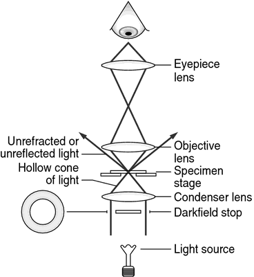
Microscope Types (with labeled diagrams) and Functions Phase-contrast microscope labeled diagram Phase-contrast microscope functions: Its applications areas include In cases where the specimen is colorless and is very tiny In biology to conduct cellular level examination of microorganisms that can't be visualized using the bright field microscopy Interference Microscope

Binocular Microscope: Features and Care – David Fankhauser
Binocular Microscope Parts Quiz - PurposeGames.com This is an online quiz called Binocular Microscope Parts. There is a printable worksheet available for download here so you can take the quiz with pen and paper. Your Skills & Rank. Total Points. 0. Get started! Today's Rank--0. Today 's Points. One of us! Game Points. 16. You need to get 100% to score the 16 points available.

Multiple Choice Quiz on Compound Microscope Parts and Functions
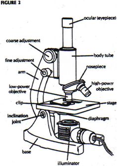
Labelled Diagram of Compound Microscope - Biology Discussion The below mentioned article provides a labelled diagram of compound microscope. Part # 1. The Stand: The stand is made up of a heavy foot which carries a curved inclinable limb or arm bearing the body tube. The foot is generally horse shoe-shaped structure (Fig. 2) which rests on table top or any other surface on which the microscope in kept.

Microscope
Compound Microscope Parts, Functions, and Labeled Diagram Common compound microscope parts include: Eyepiece (ocular lens) with or without Pointer: The part that is looked through at the top of the compound microscope. Eyepieces typically have a magnification between 5x & 30x. Monocular or Binocular Head: Structural support that holds & connects the eyepieces to the objective lenses.

File:Labelledmicroscope.gif - Wikimedia Commons
› z2 › Morphologyuni-tuebingen.de the , . of and to in a is " for on that ) ( with was as it by be : 's are at this from you or i an he have ' not - which his will has but we they all their were can ; one also the

Compound Microscope
› books › NBK310485Laboratory procedures for diagnosis of anthrax, and isolation ... In the case of Member States with limited resources and unable to operate at BSL3, it is pertinent to remember that B. anthracis is not highly infectious, and that humans are moderately resistant (see section 4.2.1). For diagnostic test purposes, therefore, good laboratory practice (Table 12) at all times is the important factor in carrying out the necessary tests safely. Large numbers of the ...
Swift Microscope Compound Microscopio 40x-1000x Monocular ...
› nature › articlesBrowse Articles | Nature Aug 08, 2022 · Primates that showed infantile behaviour after losing in a conflict drew consolation from their companions.

Microscope labeling
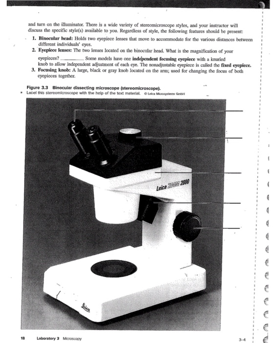
Parts of Stereo Microscope (Dissecting microscope) - labeled diagram ... Labeled part diagram of a stereo microscope Major structural parts of a stereo microscope There are three major structural parts of a stereo microscope. The viewing Head includes the upper part of the microscope, which houses the most critical optical components, including the eyepiece, objective lens, and light source of the microscope.

Draw a neat labelled diagram of a compound microscope class ...
Binocular Microscope Anatomy - Parts and Functions with a Labeled Diagram Now, I will discuss the details anatomy of the light compound microscope with the labeled diagram. Why it is called binocular: because it has two ocular lenses or an eyepiece on the head that attaches to the objective lens, this ocular lens magnifies the image produced by the objective lens. Binocular microscope parts and functions

Microscope Omano Om88 40x-1600x Clinical Compound Microscope ...
Microscope Labeling Diagram | Quizlet Unit 2 Lesson 5 - Punnett Squares and Pedigrees. 4 terms. PGFry210. Unit 2 Lesson 4 - Heredity. 9 terms. PGFry210. Upgrade to remove ads. Only $2.99/month.

Compound Microscope Parts – Labeled Diagram and their ...
Parts of a microscope with functions and labeled diagram - Microbe Notes Q. List down the 18 parts of a Microscope. 1. Ocular Lens (Eye Piece) 2. Diopter Adjustment 3. Head 4. Nose Piece 5. Objective Lens 6. Arm (Carrying Handle) 7. Mechanical Stage 8. Stage Clip 9. Aperture 10. Diaphragm 11. Condenser 12. Coarse Adjustment 13. Fine Adjustment 14. Illuminator (Light Source) 15. Stage Controls 16. Base 17.
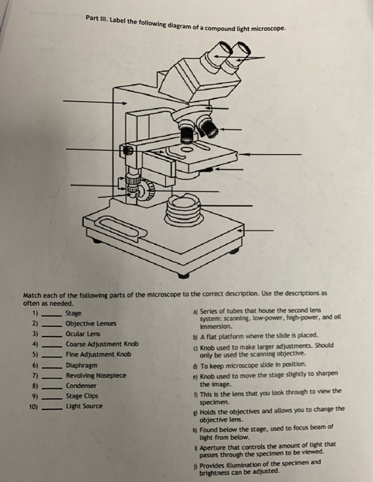
Solved Part III. Label the following diagram of a compound ...
Jual Produk Microscope Termurah dan Terlengkap Agustus 2022 ...

Stereo Compound Microscope--AmScope Supplies 40X-2000X Vet Clinical Stereo Compound Microscope B400AB

Free Microscope Drawing, Download Free Microscope Drawing png ...
Microscope Types (with labeled diagrams) and Functions

A Study of the Microscope and its Functions With a Labeled ...

monocular labeling Diagram | Quizlet

Compound Microscope Parts, Functions, and Labeled Diagram ...

Motic BA310 Digital Biological Microscope

parts of microscope with diagram

EMZ Series Zoom Stereo - Binocular and Trinocular Zoom Stereo ...

Image seen under DIY Microscope illustrate - Brainly.ph

Microscopes. (a) Binocular dissecting microscope. (b ...
Parts of a microscope with functions and labeled diagram

Microscope Parts and Functions
Instruments of Microscopy | Microbiology | | Course Hero
Trinocular Senyawa Mikroskop AmScope Perlengkapan Trinocular ...
How do microscopes operate? - Krüss laboratory equipment
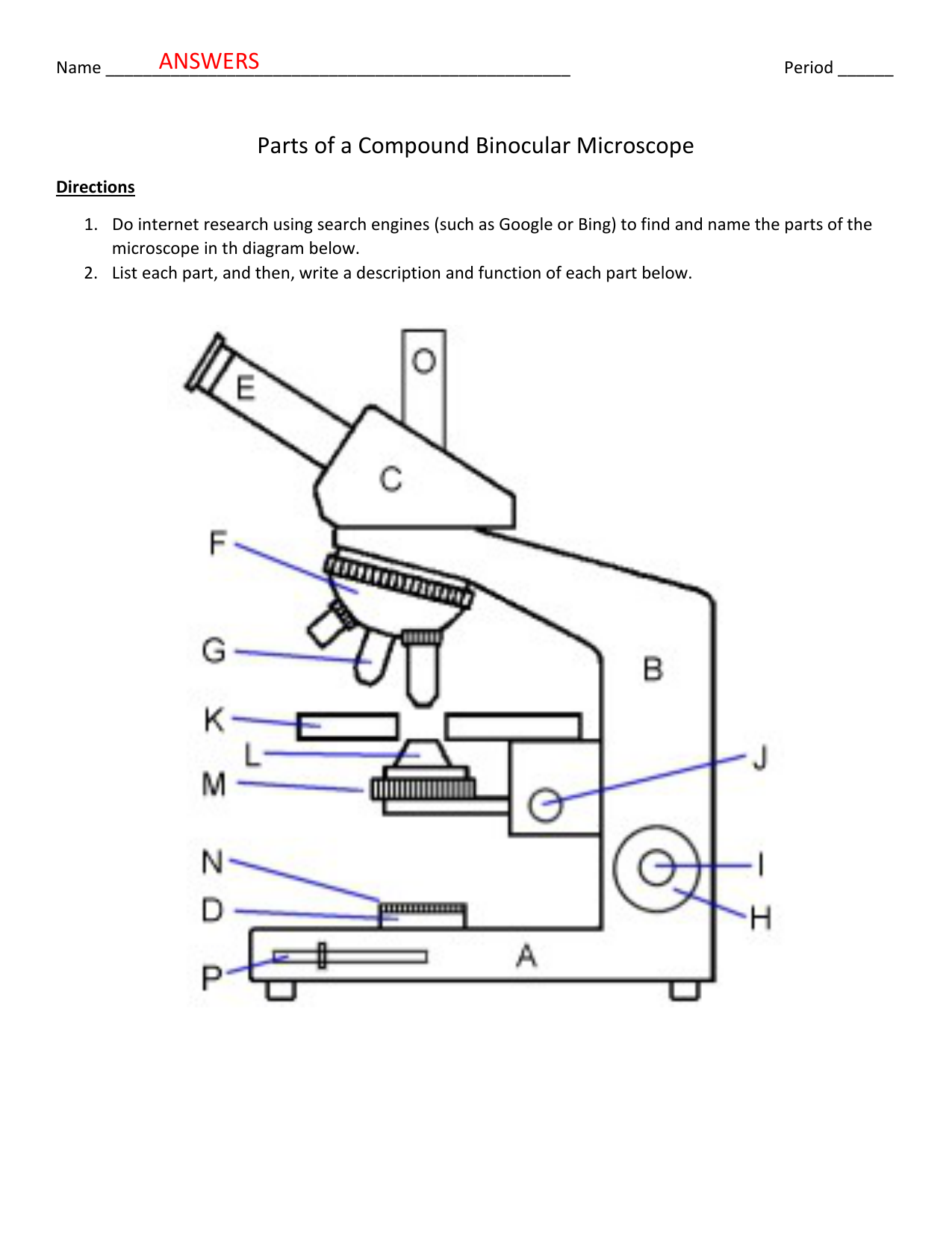
Parts of a Compound Binocular Microscope ANSWERS | Manualzz
Labelled Diagram of Microscope Parts

Diagram Of Microscope - ClipArt Best
Untitled Document
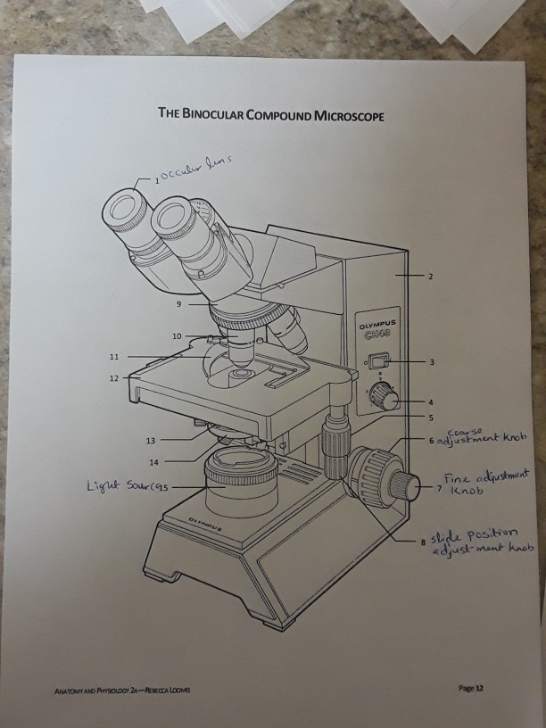
Solved THE BINOCULAR COMPOUND MICROSCOPE Cc oL 10 GHOO 12 13 ...
Binocular Microscope Anatomy - Parts and Functions with a ...

Pin page
Binocular microscope | definition of binocular microscope by ...

MICROSCOPE Drawing
Label the parts of the microscopes, and also answer | Chegg.com

Microscope Diagram Labeled, Unlabeled and Blank | Parts of a ...

Microscope With Labels Clip Art at Clker.com - vector clip ...
Microscope Diagram To Label - ClipArt Best

Diagram of a Compound Microscope

Produk Microscope | UD Berkah Abadi
Post a Comment for "45 labelled diagram of a binocular microscope"