45 how to label gel electrophoresis
A method for labeling polyacrylamide gels - Scientific Protocols Mar 24, 2011 ... Take the short plate and label on the side of the plate facing the long plate, using a laboratory permanent marker. You can write down the ... How to Read, Interpret and Analyze Gel Electrophoresis Results? How to read gel electrophoresis results? First, make clear if a gel contains any results or not. For that, put the gel carefully under the UV light and see if it contains any bands or not. In the second step, see if the gel possesses any visible contaminants like protein or RNA, or not.
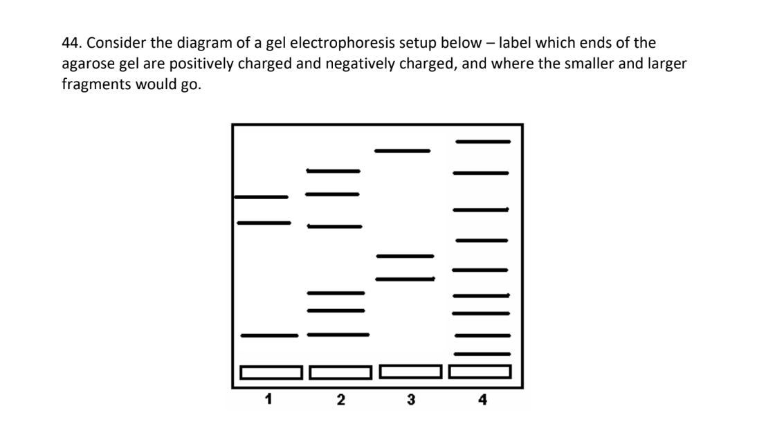
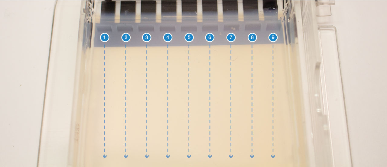
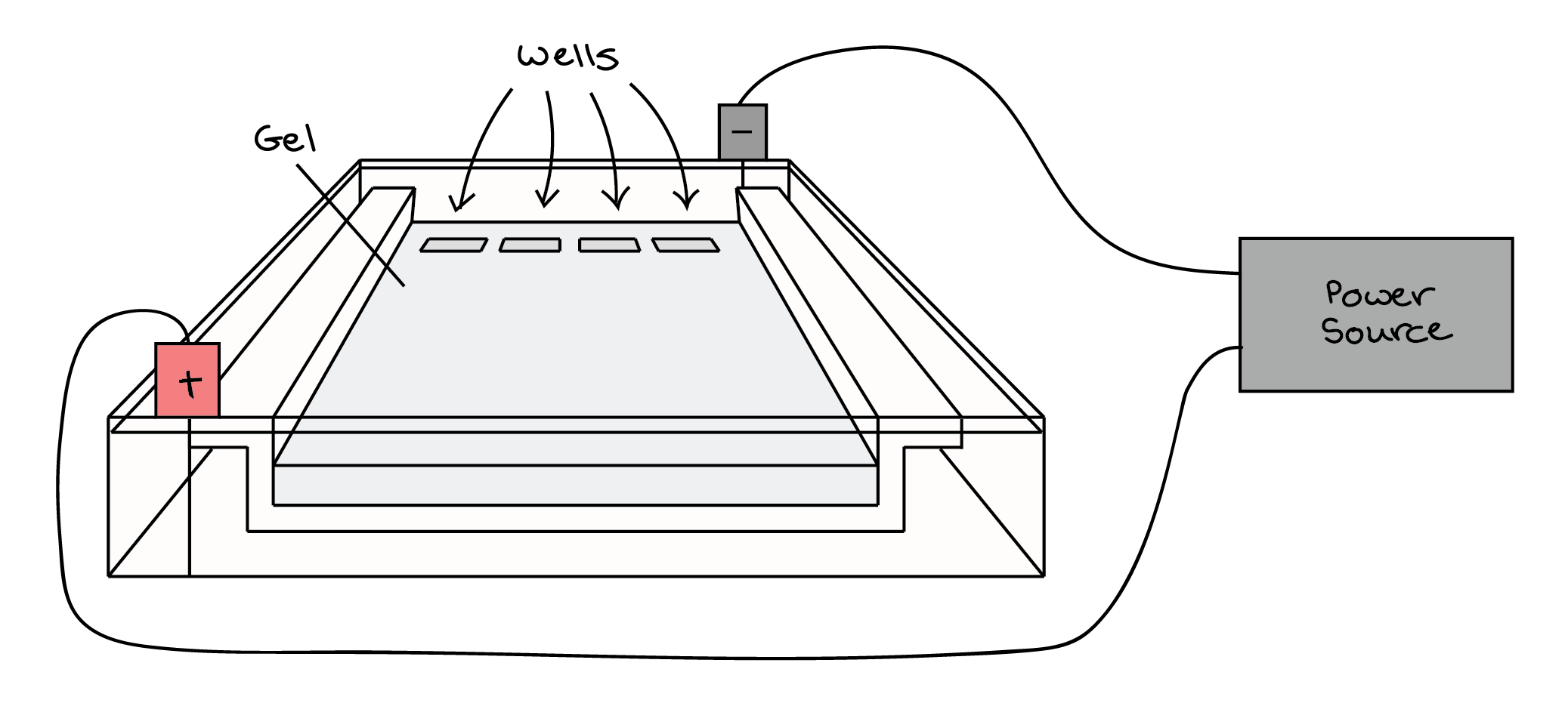
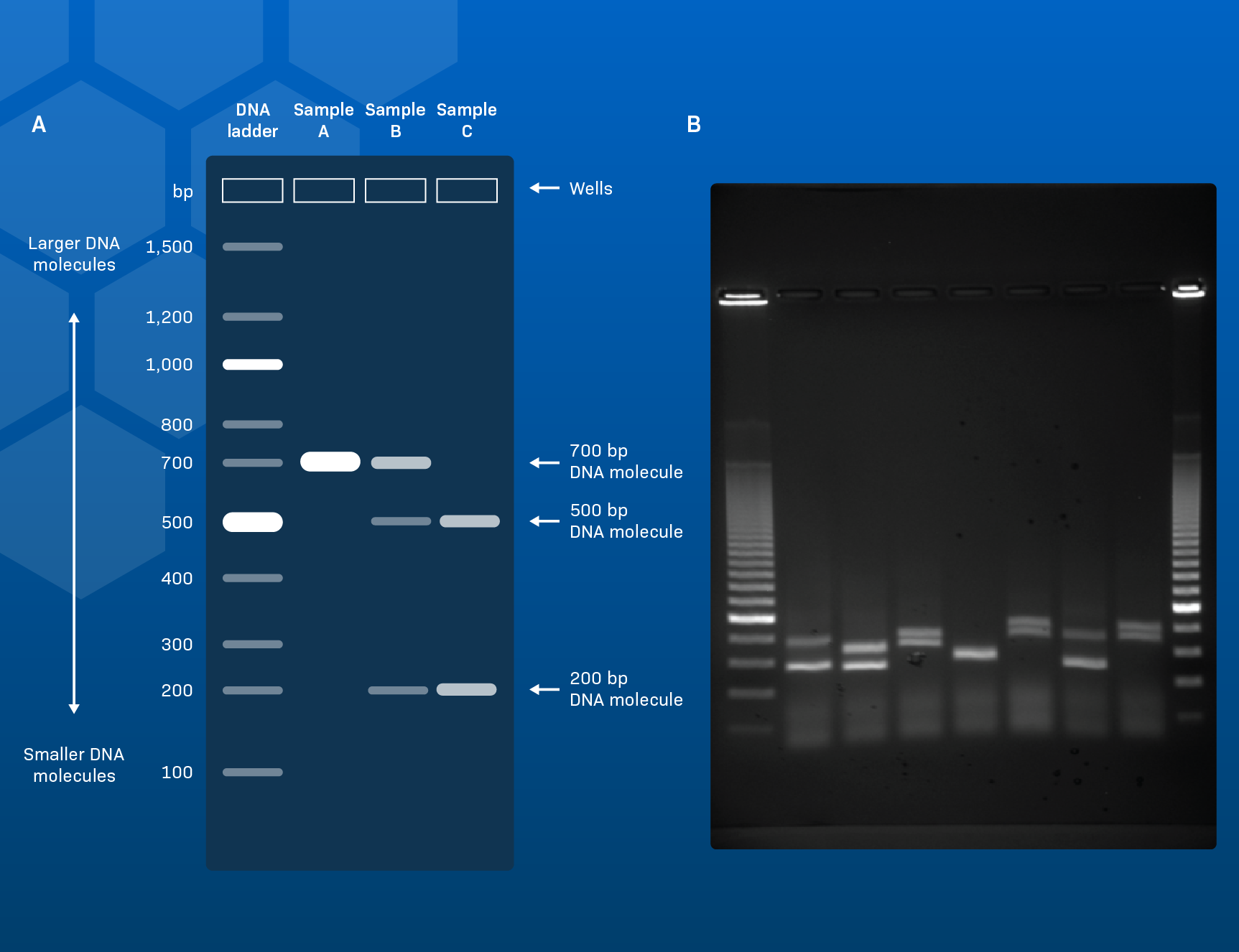
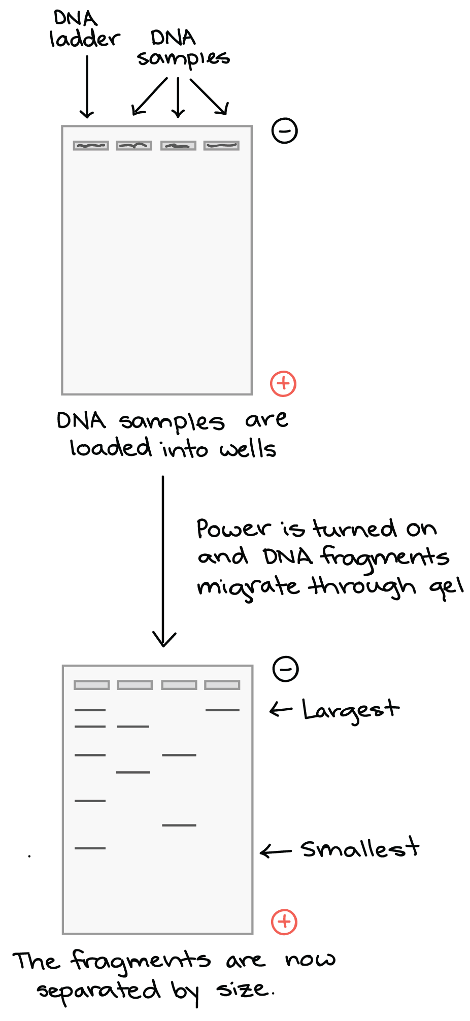
Gel electrophoresis (article) - Khan Academy Gel electrophoresis is a technique used to separate DNA fragments according to their size. DNA samples are loaded into wells (indentations) at one end of a gel, and an electric current is applied to pull them through the gel. DNA fragments are negatively charged, so they move towards the positive electrode.

How to label gel electrophoresis
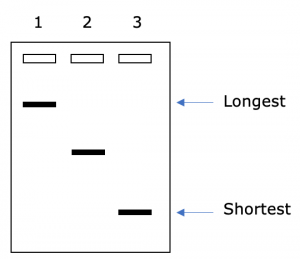
Gel electrophoresis — Science Learning Hub A solution of DNA molecules is placed in a gel. Because each DNA molecule is negatively charged, it can be pulled through the gel by an electric field. Small DNA molecules move more quickly through the gel than larger DNA molecules. The result is a series of 'bands', with each band containing DNA molecules of a particular size. › How-to-Prepare-anHow to Prepare an Electrophoresis Argarose Gel - Instructables These samples usually consist of DNA, RNA, or protein molecules. The uses of gel electrophoresis include: estimation of the size of cloned DNA, analysis of PCR products, or separation of genomic DNA. Because chemicals used in gel electrophoresis can be hazardous, no one should attempt casting a gel without basic lab safety training. What is gel electrophoresis? - YourGenome Once the DNA has migrated far enough across the gel, the electrical current is switched off and the gel is removed from the electrophoresis tank. To visualise the DNA, the gel is stained with a fluorescent dye that binds to the DNA, and is placed on an ultraviolet transilluminator which will show up the stained DNA as bright bands.
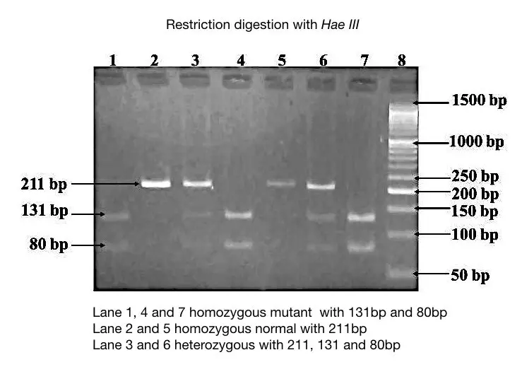
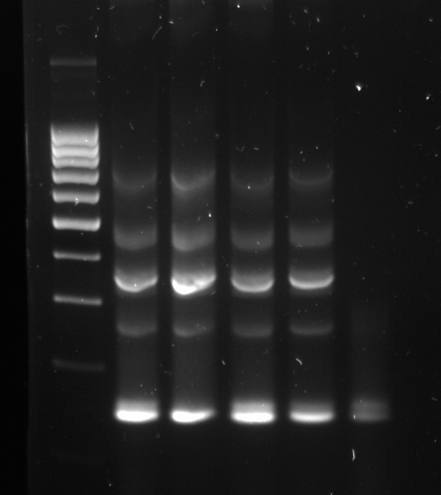
How to label gel electrophoresis. Figure legends Figure 1: Agarose gel electrophoresis (2% agarose ... Download scientific diagram | Figure legends Figure 1: Agarose gel electrophoresis (2% agarose) of PCR amplified products using species-specific PCR primer ... › intGivenchy official site Discover all the collections by Givenchy for women, men & kids and browse the maison's history and heritage researchtweet.com › gel-electrophoresis-definitionGel Electrophoresis: Definition, Principle, and Application Jul 24, 2021 · High Sensitivity Protein Gel Electrophoresis Label Compatible with Mass-Spectrometry. Biosensors (Basel) . 2020 Oct 31;10(11):160. Two-dimensional gel electrophoresis. Curr Protoc Immunol . 2005 Nov;Chapter 8:Unit 8.5. Ultrarapid Sodium Dodecyl Sulfate Polyacrylamide Mini-Gel Electrophoresis. Methods Mol Biol . 2019;1855:491-494. Part 2: Analysing and Interpreting (Agarose) Gel Electrophoresis Results The agarose gel electrophoresis is a molecular genetic technique used to separate DNA on the basis of size and charge of it. The negatively charged DNA migrates towards the positive node under the influence of the current. The results of agarose electrophoresis are affected by some of the factors enlisted below, The concentration of gel
en.wikipedia.org › wiki › Capillary_electrophoresisCapillary electrophoresis - Wikipedia Capillary electrophoresis (CE) is a family of electrokinetic separation methods performed in submillimeter diameter capillaries and in micro- and nanofluidic channels.Very often, CE refers to capillary zone electrophoresis (CZE), but other electrophoretic techniques including capillary gel electrophoresis (CGE), capillary isoelectric focusing (CIEF), capillary isotachophoresis and micellar ... PDF Gel Electrophoresis: How Does It Work - Purdue University Simply put, gel electrophoresis uses positive and negative charges to separate charged particles. Particles can be positively charged, negatively charged, or neutral. Charged particles are attracted to opposite charges: InDesign Labeling / Annotating PCR Gel Pictures - YouTube In this tutorial we will learn how to annotate Agarose Gel Pictures with Adobe InDesign CS5. I see people often labeling pictures in Photoshop and I can't re... Annotating Gels, Aligning text, and saving to a file - YouTube Short video on how to label your gels, align your text, and save to a file for later recall.
Lab 4: Gel Electrophoresis - Vanderbilt University Gel electrophoresis is a method of separating DNA fragments by movement through a Jello-like ... Google Slides or Preview to label each gel. lab-label.com › blogs › mainGel Electrophoresis - Everything You Need To Know - The Lab Label Nov 18, 2021 · After the gel electrophoresis is done, the fragments can be further used for purification or for further use in PCR. All in all, agarose gel electrophoresis is a powerful method for separating and analysing DNA and RNA fragments based on size! Method walkthrough Step 1: Making an agarose gel. The gel is made of agarose as the name of the method ... Gel Electrophoresis - Definition, Purpose and Steps - Biology Dictionary Gel electrophoresis is a procedure used to separate biological molecules by size. The separation of these molecules is achieved by placing them in a gel with small pores and creating an electric field across the gel. The molecules will move faster or slower based on their size and electric charge. Gel Electrophoresis Overview Gel electrophoresis (video) | Khan Academy 7 years ago. Yes the charge does affect the kinetics. In terms of DNA gel electrphoresis, DNA molecs already have a constant mass to charge ratio because every nucleotide has a -ve phosphate group. However for proteins (made up of different amino acids with varying charge) the charge can affect the kinetics.
Agarose gel electrophoresis of labeled DNA in which the same gel is... Following labeling with LabelIT CX-rhodamine, fluorescent DNA is visible in the absence of ethidium bromide staining due to the covalently attached rhodamine ( ...
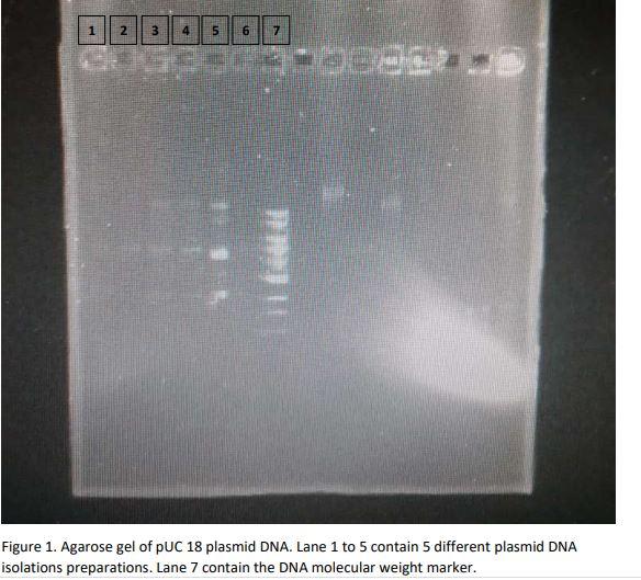
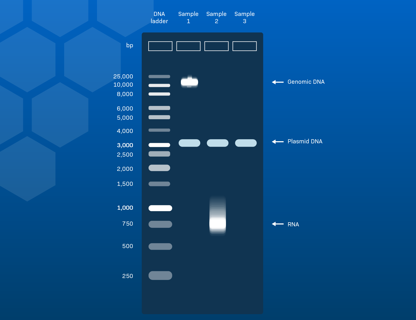
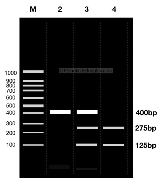
How to Interpret DNA Gel Electrophoresis Results - GoldBio During gel electrophoresis, you may have to load uncut plasmid DNA, digested DNA fragment, PCR product, and probably genomic DNA that you use as a PCR template into the wells. Your digested DNA fragment is a digested PCR product. The next step is to identify those bands to figure out which one to cut. Gel Electrophoresis. Lane 1: DNA Ladder.
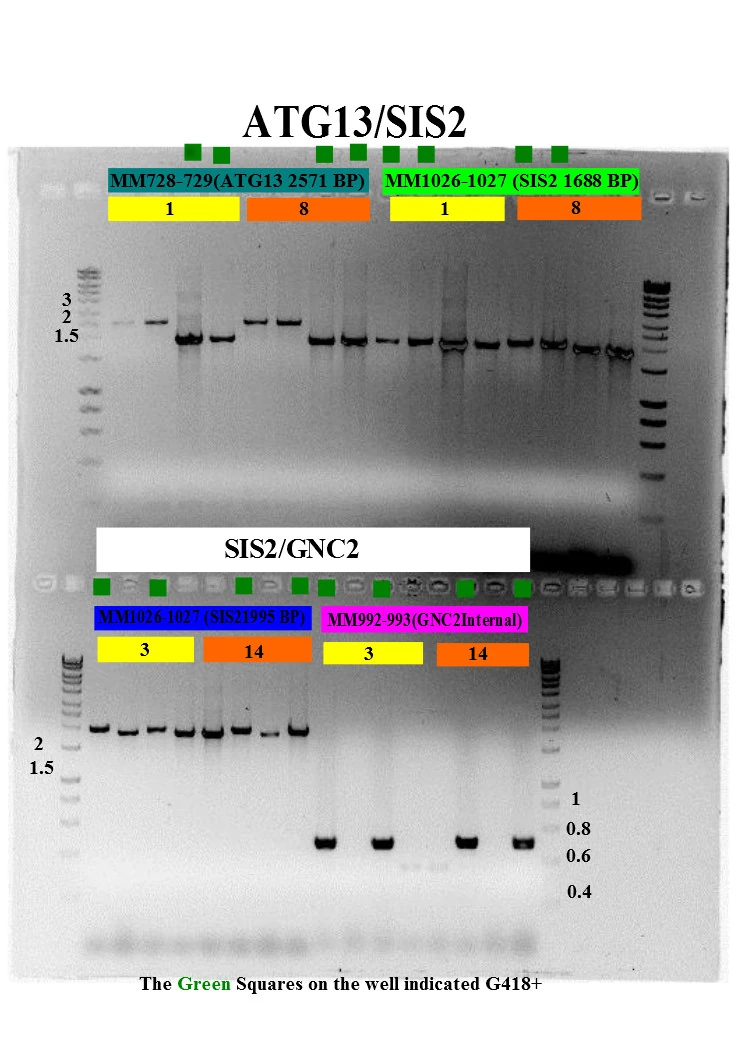
Annotating A Gel | Get Your Science On Wiki | Fandom 1.In Inkscape import your gel file and adjust the size of your picture to fit the page out line (increase zoom if needed). 2. Add in the significant ladder measurements. (On Mark's Lab area wall or just ask Mark!) 3. Create color coded rectangles to give a background for the following text. 4. Label what you PCR'd and gelled (kind of like a title).
3 min: Annotating a Gel - YouTube Mar 9, 2021 ... Your browser can't play this video. Learn more. Switch camera.
High Sensitivity Protein Gel Electrophoresis Label Compatible with ... Oct 31, 2020 ... Sodium dodecyl sulfate polyacrylamide gel electrophoresis (SDS-PAGE) is a widely utilized technique for macromolecule and protein analysis.
PDF Electrophoresis GEL and Liquid Disposal - Ohio State University electrophoresis. This electrophoresis process utilizes an organic fluorescence dye or an inorganic stain to stain the nucleic acids or proteins in a gel. These gels are typically agarose-based or polyacrylamide-based. There are a number of different protocols and dyes used in the preparation and use of electrophoresis gels.
How to label Gel electrophoresis pictures for thesis and research ... @Gelelectrophoresis @Pictures_labelling @Research_articles @Thesis @Labelling @Pictureslabellinging_in_paint @ Image_labellingHello, This video is a complete...
zlab.bio › guide-design-resourcesGuide design resources — Zhang Lab Thank you to the thousands of users who visited our guide design tool over the past five years. We recently shut down crispr.mit.edu, but there are many other guide design tools available that we hope you will find helpful.
3 Ways to Read Gel Electrophoresis Bands - wikiHow Hold a UV light up to the gel sheet to reveal results when using a UV-based dye. With your gel sheet in front of you, find the switch on a tube of UV light to turn it on. Hold the UV light 8-16 inches (20-41 cm) away from the gel sheet. Illuminate the DNA samples with the UV light to activate the dye and read the results.
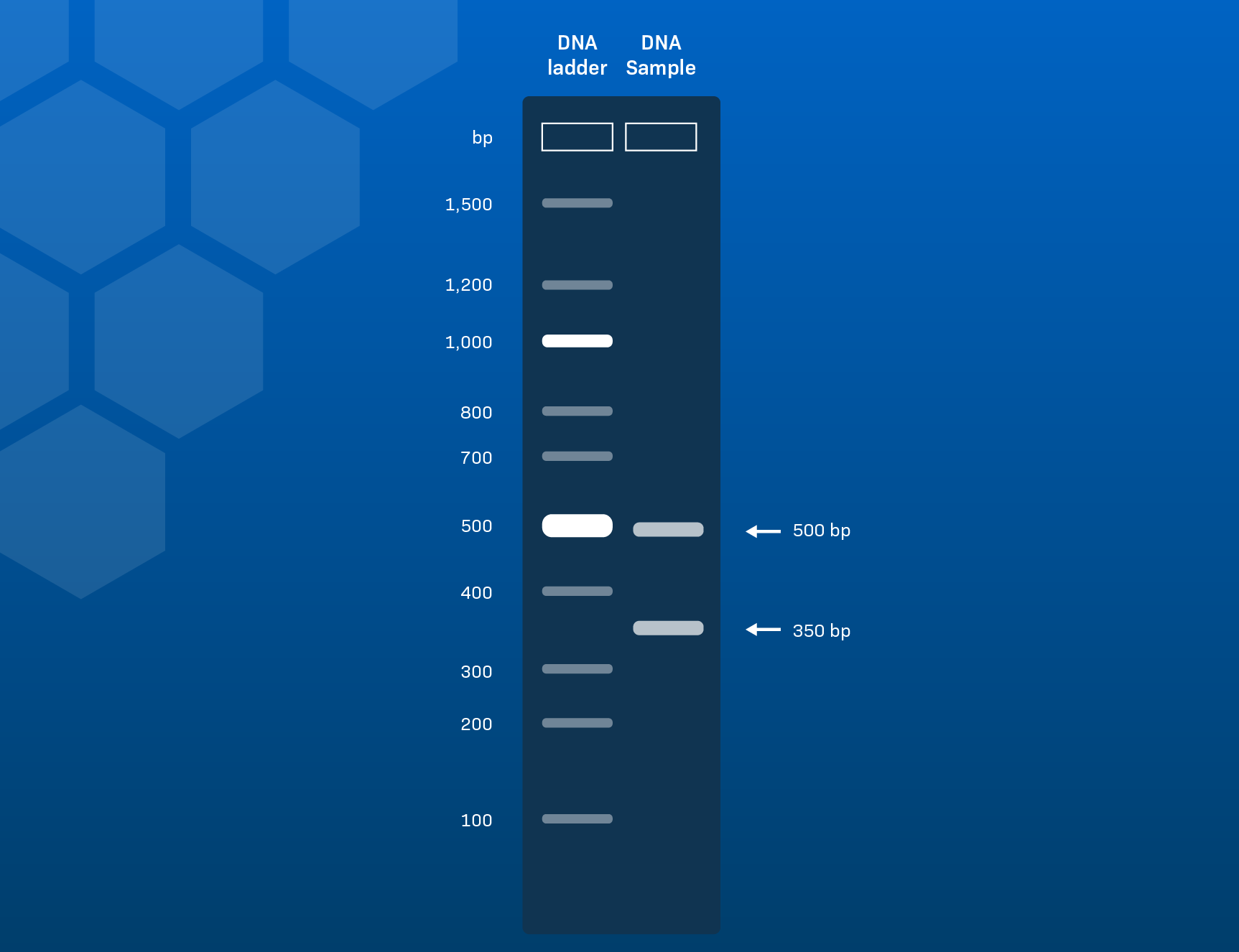
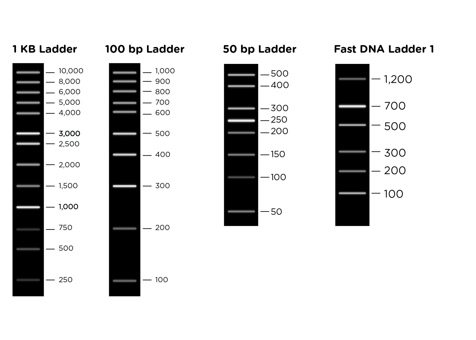
How to Interpret Agarose Gel Data: The basics - LabXchange Jun 27, 2021 ... Agarose gel electrophoresis can be used to determine how large the DNA fragment(s) in a given sample is/are. The DNA molecular weight marker, or ...
dnalc.cshl.edu › gelelectrophoresis"Gel Electrophoresis" Biology Animation Library - CSHL DNA ... In the early days of DNA manipulation, DNA fragments were laboriously separated by gravity. In the 1970s, the powerful tool of DNA gel electrophoresis was developed. This process uses electricity to separate DNA fragments by size as they migrate through a gel matrix. This animation is also available as VIDEO.
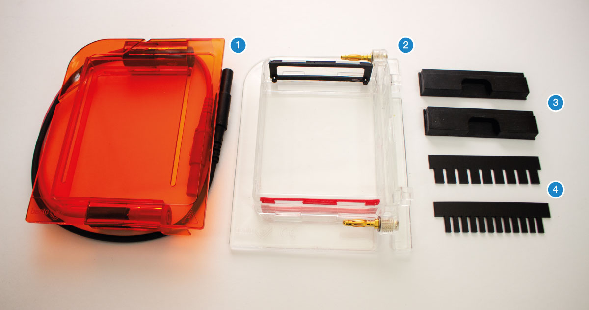
Gel Electrophoresis - Everything You Need To Know | The Lab Label Step 1: Making and agarose gel. Step 2: Setting up the power box. Step 3: Loading the samples and the molecular weight ladder. Step 4: Running the gel. Step 5: Staining the gel and analysing the gel. Tips and tricks to get the best gel electrophoresis results. The preperation and concentration of the agarose gel.
How To Label Gel Electrophoresis Images - A Strategy For The ... They are the result of gel electrophoresis, which is a common method to. Do not tackle the problem of recognizing and analyzing the labels of gel images. 1.take your jpg or png file of your gel and open it with a photo editing program (gimp). This tutorial is all about how to quickly edit & label pcr gel image using imagej software.
1.12: Restriction Digest with Gel Electrophorisis Use a Sharpie to label the top and side of 3 clean microfuge tubes A B C and your group name. Follow the reagent table below and dispense the proper amounts of reagents to the labeled tubes. Use new tips for different reagents. Add reagents to the solution at bottom of tube. Always check that your pipet tip is empty after dispensing the reagent.
ImageJ for Editing & Labelling PCR Gel Image | Biotechnology This Tutorial is all about how to quickly Edit & Label PCR Gel Image Using ImageJ software. Presented by - Elvis SamuelJoin Our Telegram Channel for free Sof...
What is gel electrophoresis? - YourGenome Once the DNA has migrated far enough across the gel, the electrical current is switched off and the gel is removed from the electrophoresis tank. To visualise the DNA, the gel is stained with a fluorescent dye that binds to the DNA, and is placed on an ultraviolet transilluminator which will show up the stained DNA as bright bands.
› How-to-Prepare-anHow to Prepare an Electrophoresis Argarose Gel - Instructables These samples usually consist of DNA, RNA, or protein molecules. The uses of gel electrophoresis include: estimation of the size of cloned DNA, analysis of PCR products, or separation of genomic DNA. Because chemicals used in gel electrophoresis can be hazardous, no one should attempt casting a gel without basic lab safety training.
Gel electrophoresis — Science Learning Hub A solution of DNA molecules is placed in a gel. Because each DNA molecule is negatively charged, it can be pulled through the gel by an electric field. Small DNA molecules move more quickly through the gel than larger DNA molecules. The result is a series of 'bands', with each band containing DNA molecules of a particular size.











-03.1624863098981-21c50c071da0c535da2ef0fcfb6c1e21.png)


Post a Comment for "45 how to label gel electrophoresis"