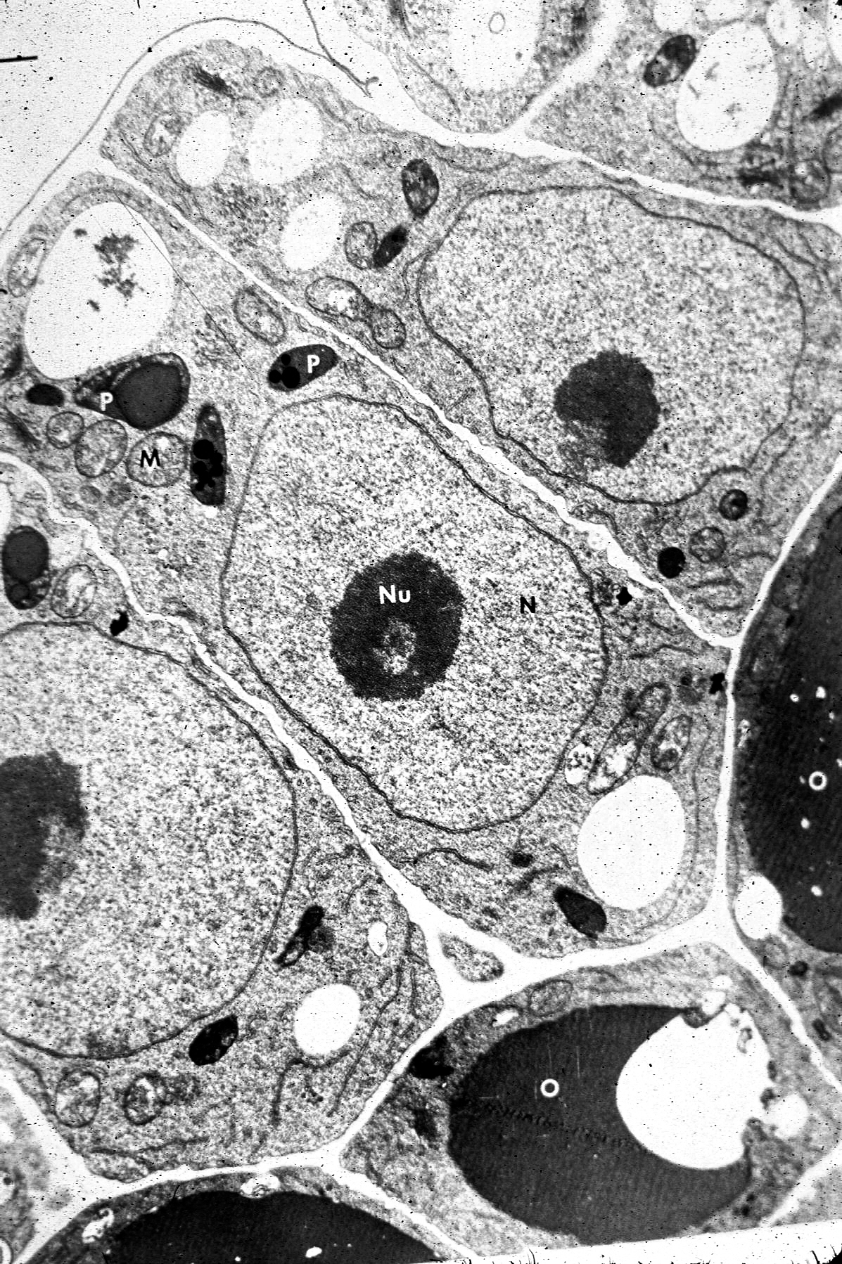
43 provide the labels for the electron micrograph
Academia.edu - Cambridge International AS and A Level Biology ... VerkkoEnzymes provide active site where the reaction can take place 2. Molecule on which an enzyme specifically acts is called the substrate 3. Binding of substrate brings about temporary change in enzyme shape known as induced fit 4. Chemical reaction occurs and substrate is changed 5. Scanning electron micrograph of D. thermolithotrophum BSA T VerkkoDownload scientific diagram | Scanning electron micrograph of D. thermolithotrophum BSA T from publication: Complete genome sequence of the thermophilic sulfur-reducer Desulfurobacterium ...
Submission guidelines | Scientific Reports - Nature VerkkoYou should provide sequencing or functional data that validates the identity of their biological constructs (plasmids, fusion proteins, site-directed mutants, etc.) either in the manuscript text ...
Provide the labels for the electron micrograph
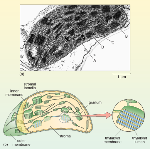
Chloroplast - Wikipedia VerkkoA chloroplast (/ ˈ k l ɔːr ə ˌ p l æ s t,-p l ɑː s t /) is a type of membrane-bound organelle known as a plastid that conducts photosynthesis mostly in plant and algal cells.The photosynthetic pigment chlorophyll captures the energy from sunlight, converts it, and stores it in the energy-storage molecules ATP and NADPH while freeing oxygen from … Electron microscope - Wikipedia VerkkoAn electron microscope is a microscope that uses a beam of accelerated electrons as a source of illumination. As the wavelength of an electron can be up to 100,000 times shorter than that of visible light photons, electron microscopes have a higher resolving power than light microscopes and can reveal the structure of smaller objects. A … en.wikipedia.org › wiki › Electron_microscopeElectron microscope - Wikipedia An electron microscope is a microscope that uses a beam of accelerated electrons as a source of illumination. As the wavelength of an electron can be up to 100,000 times shorter than that of visible light photons, electron microscopes have a higher resolving power than light microscopes and can reveal the structure of smaller objects.
Provide the labels for the electron micrograph. › cemf › whatisemWhat is Electron Microscopy? - UMass Chan Medical School Electron microscopy is used in conjunction with a variety of ancillary techniques (e.g. thin sectioning, immuno-labeling, negative staining) to answer specific questions. EM images provide key information on the structural basis of cell function and of cell disease. en.wikipedia.org › wiki › ChloroplastChloroplast - Wikipedia A chloroplast (/ ˈ k l ɔːr ə ˌ p l æ s t,-p l ɑː s t /) is a type of membrane-bound organelle known as a plastid that conducts photosynthesis mostly in plant and algal cells.The photosynthetic pigment chlorophyll captures the energy from sunlight, converts it, and stores it in the energy-storage molecules ATP and NADPH while freeing oxygen from water in the cells. Efficient and stable emission of warm-white light from lead Verkko7. marrask. 2018 · Nature - After alloying with metal cations, a lead-free halide double perovskite shows stable performance and remarkably efficient white-light emission, with possible applications in lighting and... en.wikipedia.org › wiki › Scanning_electron_microscopeScanning electron microscope - Wikipedia History. An account of the early history of scanning electron microscopy has been presented by McMullan. Although Max Knoll produced a photo with a 50 mm object-field-width showing channeling contrast by the use of an electron beam scanner, it was Manfred von Ardenne who in 1937 invented a microscope with high resolution by scanning a very small raster with a demagnified and finely focused ...
Transmission electron microscopy DNA sequencing - Wikipedia VerkkoTransmission electron microscopy DNA sequencing is a single-molecule sequencing technology that uses transmission electron microscopy techniques. The method was conceived and developed in the 1960s and 70s, but lost favor when the extent of damage to the sample was recognized. In order for DNA to be clearly visualized under an … Scanning electron microscope - Wikipedia VerkkoHistory. An account of the early history of scanning electron microscopy has been presented by McMullan. Although Max Knoll produced a photo with a 50 mm object-field-width showing channeling contrast by the use of an electron beam scanner, it was Manfred von Ardenne who in 1937 invented a microscope with high resolution by … › articles › nmat4526The surface science of nanocrystals | Nature Materials Jan 22, 2016 · Shown here is a scanning transmission electron micrograph of a cross-section of the composite. d , Sintering of NCs into a continuous film allows the production of functional solids with large ... › pages › ijoInternational Journal of Oncology - Spandidos Publications If labels cannot fit on the 17‐cm‐wide page unless the font size is smaller than 8 points, the figure must be split into several parts. Font style and appearance; Labels must be saved using standard fonts (Times New Roman, Times, Arial, Helvetica or Symbol font). The labels should be of the same font and size in all figures.
Immuno-gold electron micrograph of choroid plexus epithelial … VerkkoDownload scientific diagram | Immuno-gold electron micrograph of choroid plexus epithelial cell from a wild type mouse after cisternal kaolin injection. There is gold labelling (arrows) of AQP1 ... Clearance of senescent glial cells prevents tau-dependent Verkko19. syysk. 2018 · In a mouse model of tau-dependent neurodegenerative disease, the clearance of senescent glial cells prevents the degeneration of cortical and hippocampal neurons and preserves cognitive function. › microscopy › enZEISS LSM 900 with Airyscan 2 ZEN microscopy software puts a wealth of helpers at your command to achieve reproducible results in the shortest possible time. AI Sample Finder helps you quickly find regions of interest, leaving more time for experiments. Smart Setup supports you in applying best imaging settings for your fluorescent labels. en.wikipedia.org › wiki › Electron_microscopeElectron microscope - Wikipedia An electron microscope is a microscope that uses a beam of accelerated electrons as a source of illumination. As the wavelength of an electron can be up to 100,000 times shorter than that of visible light photons, electron microscopes have a higher resolving power than light microscopes and can reveal the structure of smaller objects.
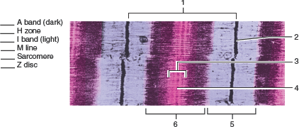
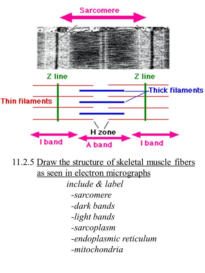
Ch 19 Skeletal Muscle Structure Flashcards | Chegg.com
Electron microscope - Wikipedia VerkkoAn electron microscope is a microscope that uses a beam of accelerated electrons as a source of illumination. As the wavelength of an electron can be up to 100,000 times shorter than that of visible light photons, electron microscopes have a higher resolving power than light microscopes and can reveal the structure of smaller objects. A …

plant cell label electron micrograph Diagram | Quizlet
Chloroplast - Wikipedia VerkkoA chloroplast (/ ˈ k l ɔːr ə ˌ p l æ s t,-p l ɑː s t /) is a type of membrane-bound organelle known as a plastid that conducts photosynthesis mostly in plant and algal cells.The photosynthetic pigment chlorophyll captures the energy from sunlight, converts it, and stores it in the energy-storage molecules ATP and NADPH while freeing oxygen from …
Figure 19.1 Srructures found in skeletal muscle fibers (cells ...

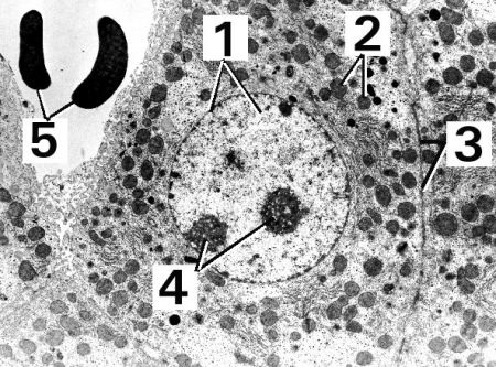
AICE Biology Chapter 1: Animal Cell Electron Micrograph ...

Transmission Electron Micrograph of transfected HL-1 cells ...
Figure 19.1 Srructures found in skeletal muscle fibers (cells ...
1.2 -Assignment - BIOLOGY4IBDP

Transmission electron microscope (TEM) micrograph showing the ...
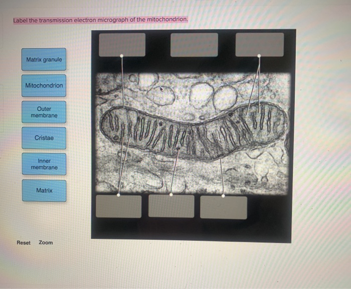
Solved Label the transmission electron micrograph of the ...

Electron micrograph of an asymmetric synapse from dissociated ...

Skeletal Muscle EM
Figure 19.1 Srructures found in skeletal muscle fibers (cells ...
What is a diagram of a plant and animal cell under an ...

Chapter 14 & 15 Flashcards Flashcards | Quizlet

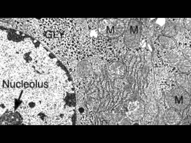
Transmission electron micrograph of cells infected with ...

Figure, Transmission Electron Micrograph of Rough Endoplasmic ...
2.3.3 Identify structures from electron micrographs of liver ...

Lung Tissue | BioNinja

1.2 Skill: Interpretation of electron micrographs
A tour of the cell: View as single page

Solved Mitochondrion Nucleus Vesicle Peroxisome | Chegg.com

how to draw chloroplast | how to draw electron micrograph of chloroplast step by step

exocrine cell of pancreas electron micrograph labelling ...

Transmission electron micrograph of a gold-labelled Lowicryl ...

1,852 Electron Micrograph Images, Stock Photos & Vectors ...

11.2 Muscles and Movement | BioNinja
Figure 19.1 Srructures found in skeletal muscle fibers (cells ...
2.2.1 Draw a generalized prokaryotic cell as seen in electron ...
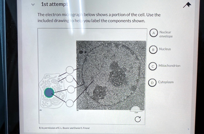
SOLVED: 1st attempt The electron micrograph below shows ...
Electron Micrograph
Biology 130 Lab 2 - Electron Micrographs
IB Biology Notes - 8.1 Cell respiration
Electron micrographs of chicken pectoralis major muscle ...

Transmission electron microscope (TEM) micrograph showing the ...

Labeling the Cell Flashcards | Quizlet

Antibody Labeling - an overview | ScienceDirect Topics
9700 QR Dynamic Papers Biology al Cambridge

Cellular portraits — Opuntia Visual
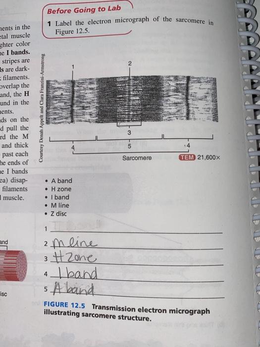
Solved Before Going to Lab Label the electron micrograph of ...
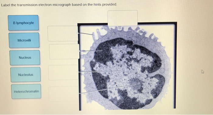
Solved Label the transmission electron micrograph based on ...
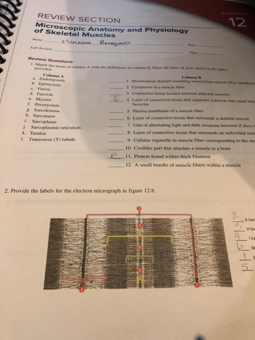
Solved 12 REVIEW SECTION Microscop of Skeletal Muscles copic ...
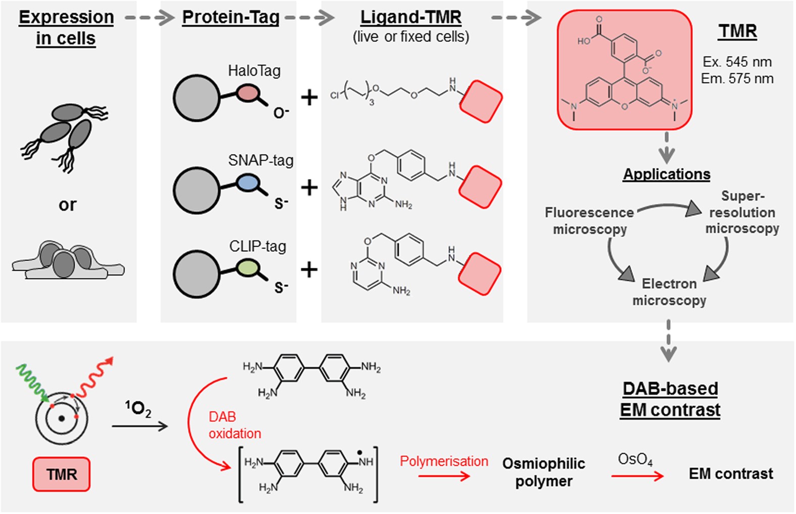
Self-labelling enzymes as universal tags for fluorescence ...

Chapter 14 & 15 Flashcards Flashcards | Quizlet
Post a Comment for "43 provide the labels for the electron micrograph"