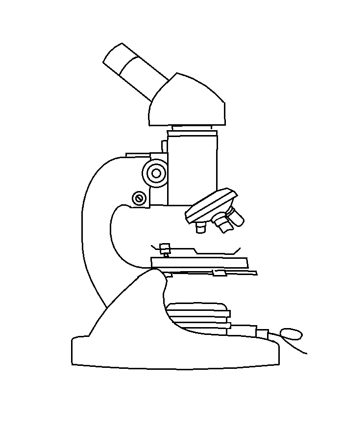
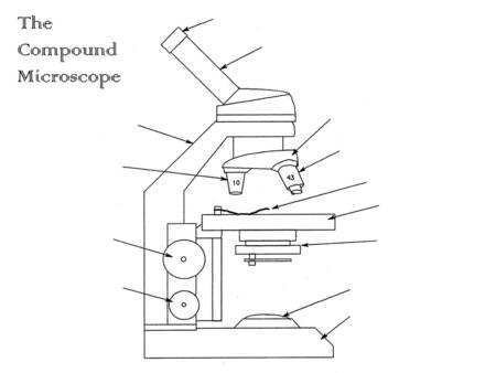
45 compound microscope diagram unlabeled
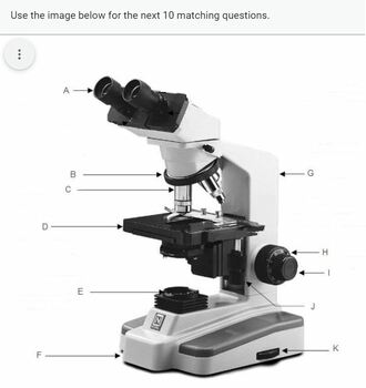
Parts of a Microscope - SmartSchool Systems Unlabeled Parts of a Microscope PDF Function of each Microscope Part 1. Eyepiece or Ocular Lens Eyepiece lens magnifies the image of the specimen. This part is also known as ocular. Most school microscopes have an eyepiece with 10X magnification. 2. Eyepiece Tube or Body Tube The tube hold the eyepiece. 3. Nosepiece Compound Microscope Labeled Diagram | Quizlet Compound Microscope Labeled + − Flashcards Learn Test Match Created by meganplocher734 Terms in this set (14) Eyepiece/Ocular lens Contains the ocular lens Body tube A hollow cylinder that holds the eyepiece. Arm Part that supports the microscope. Stage Supports the slide or specimen Coarse adjustment Knob
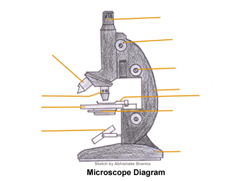
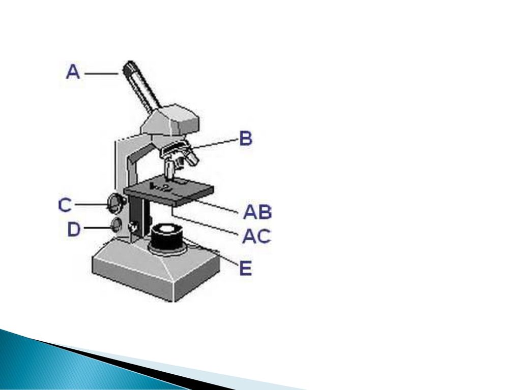
A Study of the Microscope and its Functions With a Labeled Diagram ... Here, unlabeled microscope diagrams have been provided for your perusal, which will help you practice and test your understanding of the instrument. Types of Microscopes Depending on the source of illumination, microscopes can be divided into two categories. They are:
Compound microscope diagram unlabeled
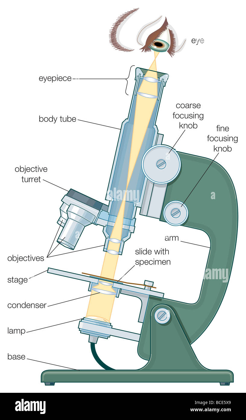
Compound Microscope- Definition, Labeled Diagram, Principle, Parts, Uses Compound microscopes have a combination of lenses that enhances both magnifying powers as well as the resolving power. The specimen or object, to be examined is usually mounted on a transparent glass slide and positioned on the specimen stage between the condenser lens and objective lens. Compound Microscope - Diagram (Parts labelled), Principle and Uses Compound Microscope Parts (Labeled diagram) Structural Components Optical Components Frequently asked Questions A compound microscope: Is used to view samples that are not visible to the naked eye Uses two types of lenses - Objective and ocular lenses Has a higher level of magnification - Typically up to 2000x Parts of a Compound Microscope - Labeled (with diagrams) Parts of a Compound Microscope - Labeled (with diagrams) A compound microscope is known as a high-power microscope that enables you to achieve a high level of magnification. Smaller specimens can be thoroughly viewed using a compound microscope. Let us take a look at the different parts of a compound microscope and understand each key component.
Compound microscope diagram unlabeled. Diagram of a Compound Microscope - Biology Discussion Diagram of a Compound Microscope Article Shared by ADVERTISEMENTS: In this article we will discuss about:- 1. Essential Parts of Compound Microscope 2. Magnification of the Image of the Object by Compound Microscope 3. Resolution Power 4. Method for Studying Microbes 5. Measurement of the Size of Objects. Essential Parts of Compound Microscope: Compound Microscope Parts - Labeled Diagram and their Functions Labeled diagram of a compound microscope Major structural parts of a compound microscope There are three major structural parts of a compound microscope. The head includes the upper part of the microscope, which houses the most critical optical components, and the eyepiece tube of the microscope. Compound Microscope Parts The three basic, structural components of a compound microscope are the head, base and arm. Base of the microscope supports the microscope and houses the illuminator. Arm connects to the base and supports the microscope head. It is also used to carry the microscope. When carrying a compound microscope always take care to lift it by both the arm ... Compound Microscope: Diagram, Parts, Working & Magnification | AESL A compound microscope is an optical instrument which uses two sets of lenses providing a high resolution and 2-dimensional image of the sample. Compound microscope is one type of optical microscope, the other type is a simple microscope. The difference between a simple and a compound microscope is that a simple microscope uses one lens whereas ...
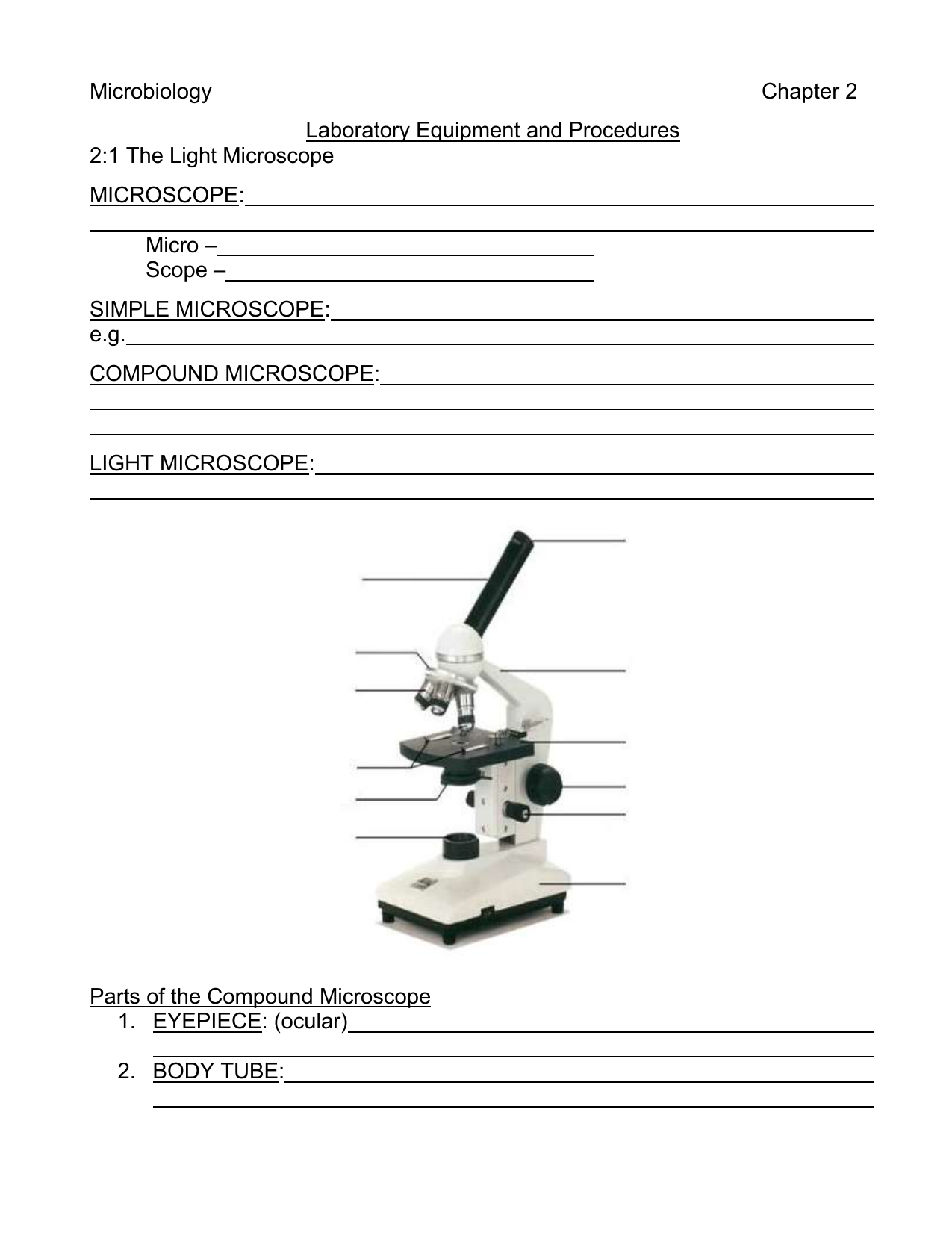
PDF AN INTRODUCTION TO THE COMPOUND MICROSCOPE - Rowan University AN INTRODUCTION TO THE COMPOUND MICROSCOPE OBJECTIVE: In this lab you will learn the basic skills needed to stain and mount wet slides. You will also learn about magnification, resolution and the parts of the compound microscope. INTRODUCTION: The light microscope can extend our ability to see detail by 1000 times, so that we can Compound Microscope Parts, Functions, and Labeled Diagram Compound Microscope Definitions for Labels Eyepiece (ocular lens) with or without Pointer: The part that is looked through at the top of the compound microscope. Eyepieces typically have a magnification between 5x & 30x. Monocular or Binocular Head: Structural support that holds & connects the eyepieces to the objective lenses. Unlabeled Microscope Diagram - Cliparts.co Unlabeled Microscope Diagram - Cliparts.co Unlabeled Microscope Diagram 59 images of Unlabeled Microscope Diagram. You can use these free cliparts for your documents, web sites, art projects or presentations. Don't forget to link to this page for attribution! Parts of a microscope with functions and labeled diagram - Microbe Notes Parts of a microscope with functions and labeled diagram September 17, 2022 by Faith Mokobi Having been constructed in the 16th Century, Microscopes have revolutionalized science with their ability to magnify small objects such as microbial cells, producing images with definitive structures that are identifiable and characterizable.
Compound Microscope Parts - Labeled Diagram and their Functions (2023) The term "compound" refers to the microscope having more than one lens. Basically, compound microscopes generate magnified images through an aligned pair of the objective lens and the ocular lens. In contrast, "simple microscopes" have only one convex lens and function more like glass magnifiers. [In this figure] Two "antique ... Compound Microscope: Definition, Diagram, Parts, Uses, Working Principle A compound microscope is defined as A microscope with a high resolution and uses two sets of lenses providing a 2-dimensional image of the sample. The term compound refers to the usage of more than one lens in the microscope. Also, the compound microscope is one of the types of optical microscopes. 16 Parts of a Compound Microscope: Diagrams and Video Once you have an understanding of the parts of the microscope it will be much easier to navigate around and begin observing your specimen, which is the fun part! The 16 core parts of a compound microscope are: Head (Body) Arm. Base. Eyepiece. Eyepiece tube. Microscopy: Intro to microscopes & how they work (article) - Khan Academy Magnification is a measure of how much larger a microscope (or set of lenses within a microscope) causes an object to appear. For instance, the light microscopes typically used in high schools and colleges magnify up to about 400 times actual size. So, something that was 1 mm wide in real life would be 400 mm wide in the microscope image.
Microscope Parts and Use Google Form Quiz
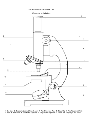
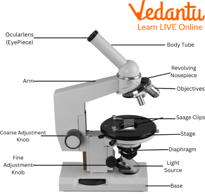
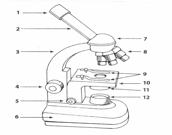
Parts of the Microscope (Labeled Diagrams) - Simple and Compound Microscope Optical Parts of a Compound Microscope. Eyepiece lens or Ocular. Mirror. Objective Lenses. Scanning Objective Lens (4x) Low Power Objective (10x) High Power Objective Lens (40x) Oil Immersion Objective Lens (100x) Specialty Objective Lenses.

Celestron LABS CM2000CF Compound Microscope
Microscope: Parts Of A Microscope With Functions And Labeled Diagram. Figure: A diagram of a microscope's components. The microscope has three basic components: the head, the base, and the arm. Head:Occasionally, the head is considered the body. It holds the optical components of the upper part of the microscope. Base:The microscope's base provides great support. It is also equipped with miniature illuminators.

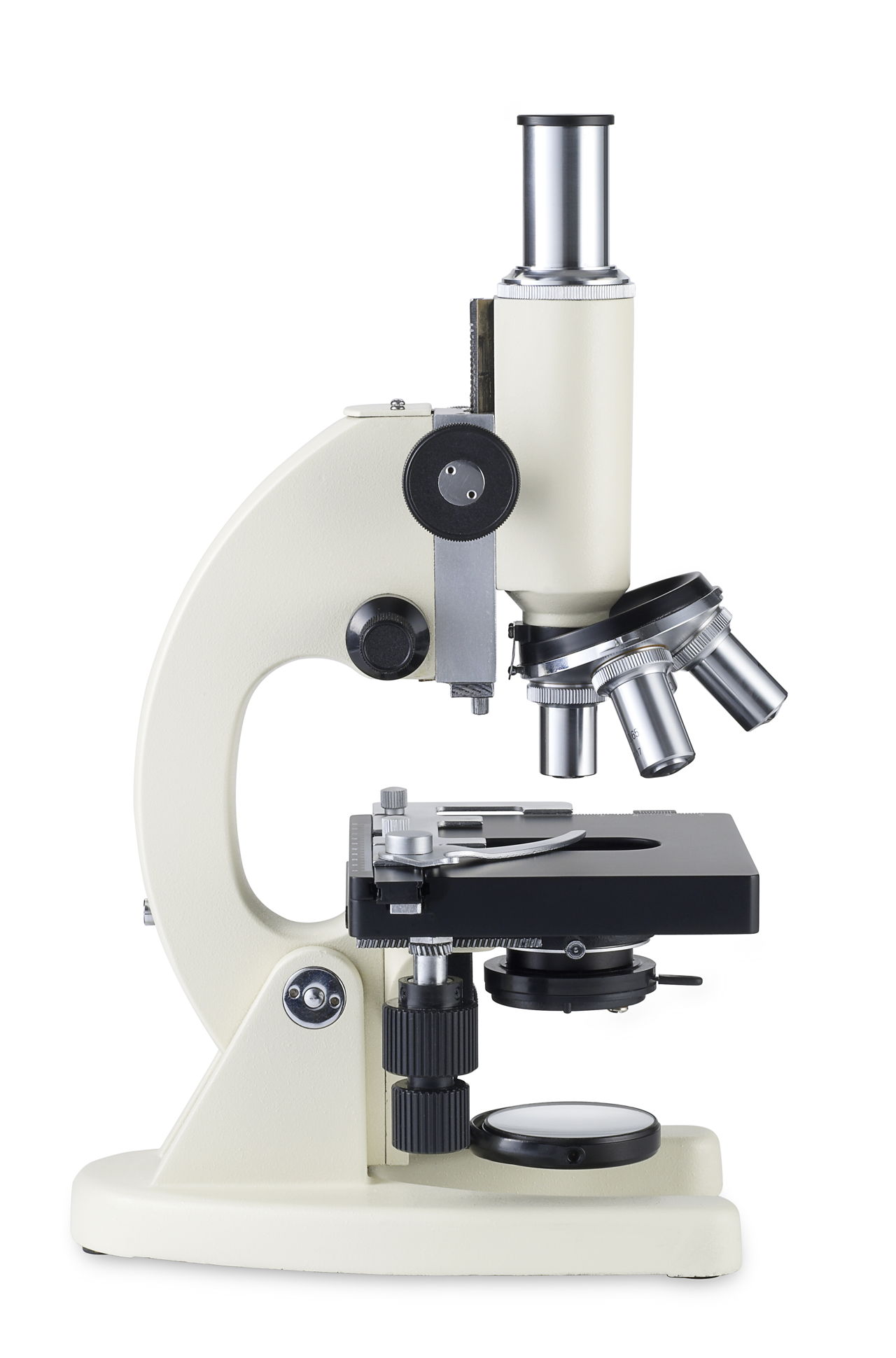
Elementary Compound Field Microscope with LED illumination ...
Microscope Parts and Functions With Labeled Diagram and Functions How does a Compound Microscope Work? Before exploring microscope parts and functions, you should probably understand that the compound light microscope is more complicated than just a microscope with more than one lens.
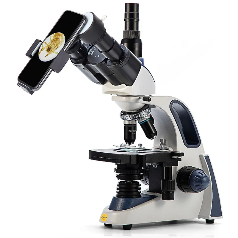
Swift-sw380t Best Price Biogical Compound Trinocular Laboratory Microscope 2500x Microscopic Portable Malaria Microscopy - Buy Compound Microscope,2 ...
Microscope Diagram Unlabeled Web microscope labeled diagram 1. It is noted first that which objective lens is in use on the microscope. Web Free Unlabeled Microscope Diagram Vector Download In Ai, Svg, Eps And Cdr. Head:occasionally, the head is considered the body. Web there are three major structural parts of a compound microscope.

Compound Microscope Parts – Labeled Diagram and their ...
Compound Microscope Parts, Function, & Diagram - Study.com Learn the compound light microscope's parts and functions by viewing a compound microscope diagram. Also, read about the uses of a compound microscope. Updated: 11/04/2021

The mechanical components of a compound microscope, Stock ...
Label the microscope — Science Learning Hub Use this interactive to identify and label the main parts of a microscope. Drag and drop the text labels onto the microscope diagram. diaphragm or iris base eye piece lens fine focus adjustment light source stage coarse focus adjustment high-power objective Download Exercise

Compound Microscope Parts – Labeled Diagram and their ...
Parts of a Compound Microscope - Labeled (with diagrams) Parts of a Compound Microscope - Labeled (with diagrams) A compound microscope is known as a high-power microscope that enables you to achieve a high level of magnification. Smaller specimens can be thoroughly viewed using a compound microscope. Let us take a look at the different parts of a compound microscope and understand each key component.

Label the microscope — Science Learning Hub
Compound Microscope - Diagram (Parts labelled), Principle and Uses Compound Microscope Parts (Labeled diagram) Structural Components Optical Components Frequently asked Questions A compound microscope: Is used to view samples that are not visible to the naked eye Uses two types of lenses - Objective and ocular lenses Has a higher level of magnification - Typically up to 2000x
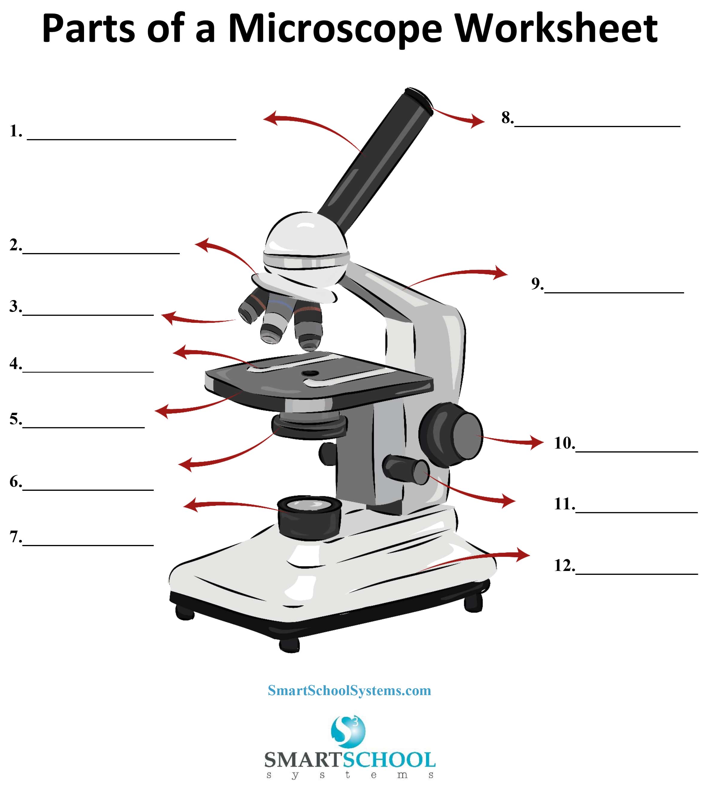
Parts of a Microscope - SmartSchool Systems
Compound Microscope- Definition, Labeled Diagram, Principle, Parts, Uses Compound microscopes have a combination of lenses that enhances both magnifying powers as well as the resolving power. The specimen or object, to be examined is usually mounted on a transparent glass slide and positioned on the specimen stage between the condenser lens and objective lens.

Parts of a Compound Microscope — Learning in Hand with Tony ...

KERN OBE 134 Compound Microscope Trinocular Achromat 4/10/40 ...
Free Microscope Drawing, Download Free Microscope Drawing png ...

Labeled Parts Of A Microscope - ClipArt Best
unlabelled diagram of the microscope - Clip Art Library

Swift SW350B 40X-2500X Magnification, Siedentopf Binocular ...
Parts Of The Microscope Worksheet - ClipArt Best
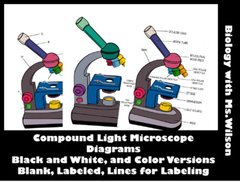
Label The Parts Of A Compound Microscope Teaching Resources | TPT

Microscope With Labels Clip Art at Clker.com - vector clip ...
(159).jpg)
Microscope Quiz: How Much You Know About Microscope Parts And ...

Microscope Diagram Labeled, Unlabeled and Blank | Parts of a ...

National Optical Model 134-CLED Compound Microscope
Components compound microscope hi-res stock photography and ...

Parts of the Microscope worksheet

Educational Dual Magnification Stereo Microscope with Built ...
Microscope Photo - Fill Online, Printable, Fillable, Blank ...

compound microscope Diagram | Quizlet

BARSKA Monocular Compound Microscope with Light, 40x, 100x ...
Microscope and Their Uses - Learn Important Terms and Concepts

Biology label part of microscope

Living Environment Course
Ch. 2 Guided Notes
Microscope Parts Quiz

Microscope Diagram Labeled, Unlabeled and Blank | Parts of a ...

Microscope Diagram (Grade 8) - Free Printable Tests and ...

Swift SW380T Siedentopf Trinocular Compound Microscope for ...
What are objectives on a microscope? How do they contribute ...

Parts of the Microscope worksheet
Line Style Vector Illustration of Microscope. Logo of ...
Microscope Lab Learn the parts which are directly used for ...
Understanding the Compound Microscope Parts and its Functions ...
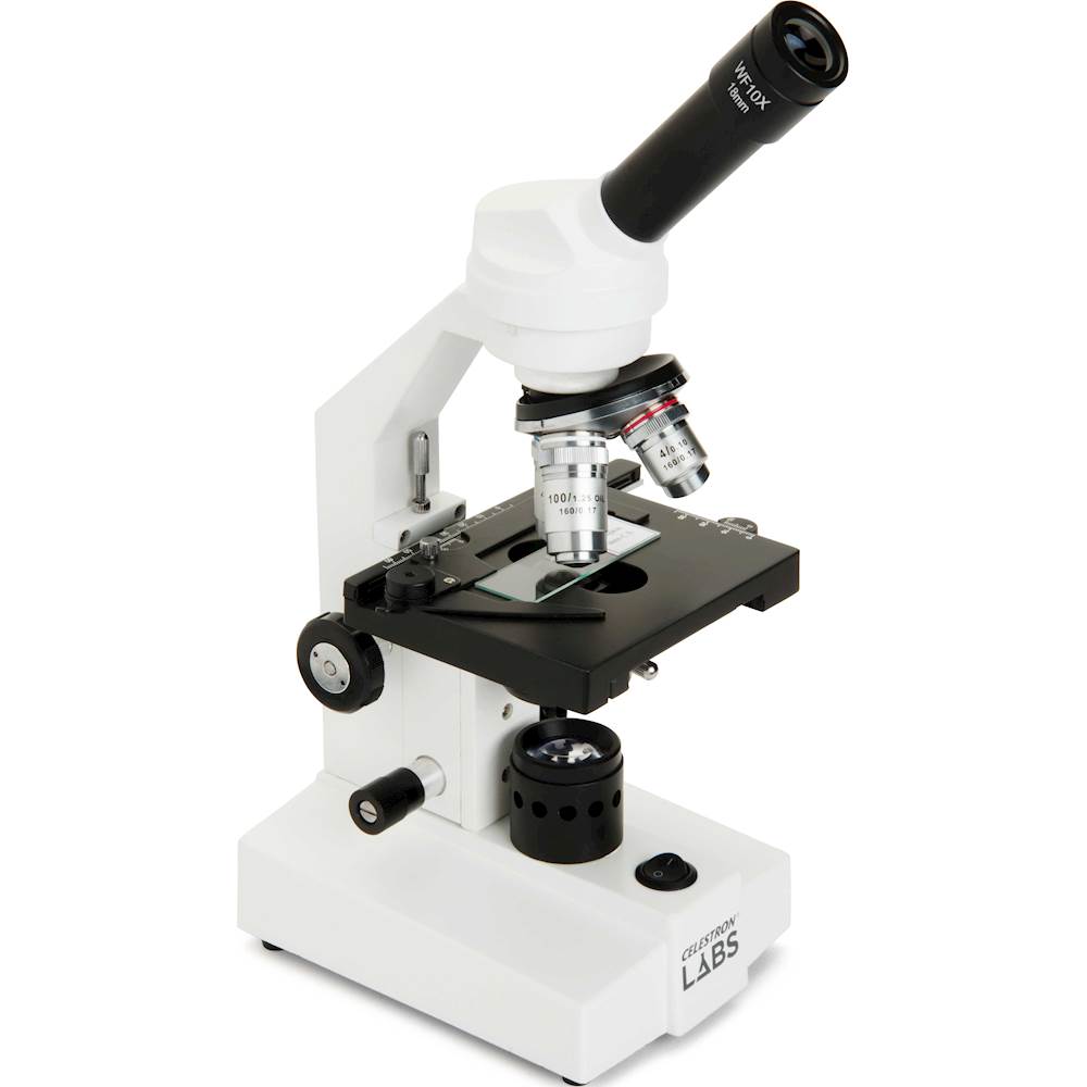
Celestron CM2000CF Compound Microscope 44130 - Best Buy

10X-60X Compact Multi-Lens Stereo Microscope with Angled Head ...

BIM151T-LED microscope with 40x, 100x, 400x and 1000x ...

Buy Compound Microscope | Compound Microscopes – Tagged ...
Parts and focusing Microscope Basics (1) Parts and focusing ...
Post a Comment for "45 compound microscope diagram unlabeled"